Review
. 2023 Dec 27;25(1):388.
doi: 10.3390/ijms25010388.

Extracellular Vesicles in Atherosclerosis: State of the Art

Affiliations
Review

Extracellular Vesicles in Atherosclerosis: State of the Art

Wioletta Olejarz et al. Int J Mol Sci. .

Abstract

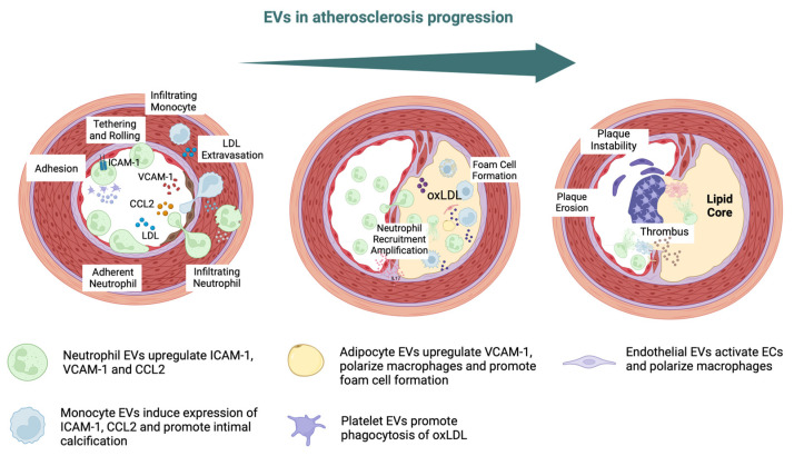
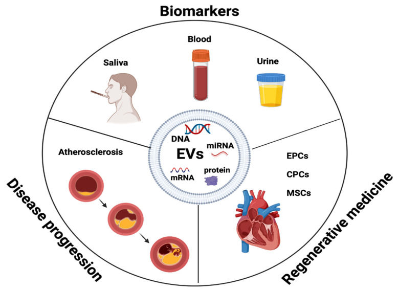
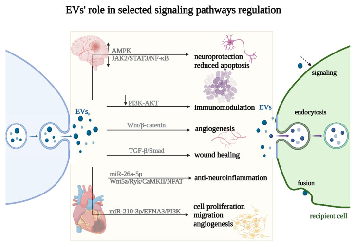
Atherosclerosis is a chronic inflammatory disease driven by lipid accumulation in the arteries, leading to narrowing and thrombosis that causes mortality. Emerging evidence has confirmed that atherosclerosis affects younger people and is involved in the majority of deaths worldwide. EVs are associated with critical steps in atherosclerosis, cholesterol metabolism, immune response, endothelial dysfunction, vascular inflammation, and remodeling. Endothelial cell-derived EVs can interact with platelets and monocytes, thereby influencing endothelial dysfunction, atherosclerotic plaque destabilization, and the formation of thrombus. EVs are potential diagnostic and prognostic biomarkers in atherosclerosis (AS) and cardiovascular disease (CVD). Importantly, EVs derived from stem/progenitor cells are essential mediators of cardiogenesis and cardioprotection and may be used in regenerative medicine and tissue engineering.

Keywords: CVD; EPC; MSC; atherosclerosis; endothelial cells; extracellular vesicles.


Conflict of interest statement

The authors declare no conflict of interest.

Figures

Figure 2
Figure 2
EVs in atherosclerosis progression. Intercellular adhesion molecule 1 (ICAM-1), vascular cell adhesion molecule-1 (VCAM-1), chemokine C–C motif chemokine ligand 2 (CCL2), low-density lipoprotein (LDL), oxidized LDL (oxLDL) [48,82].
Figure 1
Figure 1
EVs’ exterior and interior components. Major histocompatibility complex (MHC), heat shock proteins (Hsp), tumor susceptibility gene (TSG), tumor necrosis factor (TNF), TNF-related apoptosis-inducing ligand (TRAIL), Fas ligand (FasL), transferrin receptor (TfR) [28].
Figure 3
Figure 3
EVs as biomarkers in disease progression and regenerative medicine. Endothelial progenitor cells (EPCs), cardiac progenitor cells (CPCs), mesenchymal stem cells (MSCs).
Figure 4
Figure 4
miRNAs as biomarkers in AS and CVD.
Figure 5
Figure 5
Selected signaling pathways’ regulation (↑—upregulation, ↓—downregulation) by EVs. EVs can impact the recipient cell phenotype using three different uptake mechanisms: endocytosis, fusion, or signaling (marked with a dashed arrow).

Similar articles

Cited by

References

    1. Kobiyama K., Ley K. Atherosclerosis. Circ. Res. 2018;123:1118–1120. doi: 10.1161/CIRCRESAHA.118.313816. - DOI - PMC - PubMed
    1. Libby P. The changing landscape of atherosclerosis. Nature. 2021;592:524–533. doi: 10.1038/s41586-021-03392-8. - DOI - PubMed
    1. Botts S.R., Fish J.E., Howe K.L. Dysfunctional Vascular Endothelium as a Driver of Atherosclerosis: Emerging Insights into Pathogenesis and Treatment. Front. Pharmacol. 2021;12:787541. doi: 10.3389/fphar.2021.787541. - DOI - PMC - PubMed
    1. Lu L., Liu M., Sun R., Zheng Y., Zhang P. Myocardial Infarction: Symptoms and Treatments. Cell Biochem. Biophys. 2015;72:865–867. doi: 10.1007/s12013-015-0553-4. - DOI - PubMed
    1. Spacek M., Zemanek D., Hutyra M., Sluka M., Taborsky M. Vulnerable atherosclerotic plaque—A review of current concepts and advanced imaging. Biomed. Pap. Med. Fac. Univ. Palacky. Olomouc Czech Repub. 2018;162:10–17. doi: 10.5507/bp.2018.004. - DOI - PubMed