. 2023 Dec 28;25(1):446.
doi: 10.3390/ijms25010446.

Antibody Response to HERV-K and HERV-W Envelope Epitopes in Patients with Myasthenia Gravis

Affiliations

Antibody Response to HERV-K and HERV-W Envelope Epitopes in Patients with Myasthenia Gravis

Elena Rita Simula et al. Int J Mol Sci. .

Abstract

Myasthenia gravis is an antibody-mediated autoimmune neurological disorder characterized by impaired neuromuscular junction transmission, resulting in muscle weakness. Recently, the involvement of Human Endogenous Retroviruses (HERVs) in the pathophysiology of different immune-mediated and neurodegenerative diseases, such as multiple sclerosis, has been demonstrated. We aimed to investigate potential immune system involvement related to humoral responses targeting specific epitopes of HERV-K and HERV-W envelope proteins in myasthenia gravis. Myasthenia gravis patients were recruited in the Neurology Unit, while healthy controls were selected from the Blood Transfusion Center, both affiliated with AOU Sassari. Highly immunogenic antigens of HERV-K and HERV-W envelope proteins were identified using the Immune Epitope Database (IEDB) online tool. These epitopes were utilized in enzyme-linked immunosorbent assays (ELISA) to detect autoantibodies in serum directed against these sequences. The study involved 39 Healthy Donors and 47 MG patients, further categorized into subgroups based on the presence of autoantibodies: MG-AchR Ab+ (n = 17), MG-MuSK Ab+ (n = 7), double seronegative patients (MG-DSN, n = 18), MG-LRP4 Ab + (n = 4), and one patient with no antibodies data (n = 1). Our findings revealed high levels of autoantibodies in myasthenia gravis patients directed against the HERV-K-env-su(19-37), HERV-K-env-su(109-126), HERV-K-env-su(164-186), HERV-W-env(93-108), HERV-W-env(129-14), and HERV-W-env(248-262) epitopes. Notably, these results remained highly significant even when patients were subdivided into MG-AchR Ab+ and MG-DSN subgroups. Correlation analysis further revealed significant positive associations between the antibody levels against HERV-K and HERV-W families in patients, suggesting a synergistic action of the two HERVs in the pathology context since this correlation is absent in the control group. This study marks the first identification of a specific humoral response directed against defined epitopes of HERV-K and HERV-W envelope proteins in myasthenia gravis patients. These findings lay the foundation for future investigations aimed at elucidating the molecular mechanisms driving this immune response. The detection of these autoantibodies suggests the potential for novel biomarkers, especially within the MG-DSN patient subgroup, addressing the need for new biomarkers in this population.

Keywords: HERV-K; HERV-W; MG; autoantibodies; epitopes; humoral response; myasthenia gravis.


Conflict of interest statement

The authors declared no conflict of interest.

Figures
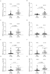
Figure 1
Figure 1
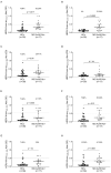
ELISA-based analysis of Abs reactivity against HERV-K-env-su- and HERV-W-env-derived peptides. Plasma samples from the entire cohort of MG patients and HCs subjects were tested against HERV-K-env-su(19–37) (A), HERV-K-env-su(109–126) (B), HERV-K-env-su(164–186) (C), HERV-K-env-su(205–226) (D), HERV-W-env(93–108) (E), HERV-W-env(129–143) (F), HERV-W-env(161–180) (G), and HERV-W-env(248–262) (H) peptides. The median and dashed lines delineate the thresholds employed to determine sample positivity. The upper section of each graph displays the p-value and the proportion of positive patients, as determined with Fisher’s exact test, “ns” indicates a non-significant result with a p-value greater than 0.05, suggesting no statistically significant differences between the compared groups.
Figure 2
Figure 2
Summary graph depicting the positivity rates among MG patients, categorized into AchR Ab+ (A) and DSN (B), in response to peptides derived from HERVs. The first bar, in deep green (A)/orange (B), represents the overall percentage of patients who tested positive for at least one of the epitopes under investigation. The bars located on the right side of the graph display the patient positivity rates for each specific peptide: three bars in light green (A)/orange (B) for HERV-K peptides, and the four bars in a darker shade of green (A)/orange (B) for HERV-W peptides.
Figure 3
Figure 3
ELISA-based analysis of Abs reactivity against HERV-K-env-su- and HERV-W-env-derived epitopes. Plasma samples from MG AchR Ab+ patients and HCs subjects were tested against HERV-K-env-su(19–37) (A), HERV-K-env-su(109–126) (B), HERV-K-env-su(164–186) (C), HERV-K-env-su(205–226) (D), HERV-W-env(93–108) (E), HERV-W-env(129–143) (F), HERV-W-env(161–180) (G), and HERV-W-env(248–262) (H) peptides. The median and dashed lines delineate the thresholds employed to determine sample positivity. The upper section of each graph displays the p-value and the proportion of positive patients, as determined with Fisher’s exact test, “ns” indicates a non-significant result with a p-value greater than 0.05, suggesting no statistically significant differences between the compared groups.
Figure 4
Figure 4
ELISA-based analysis of Abs reactivity against HERV-K-env-su and HERV-W-env-su derived peptides. Plasma samples from MG Musk Ab+ patients and HCs subjects were tested against HERV-K-env-su(19–37) (A), HERV-K-env-su(109–126) (B), HERV-K-env-su(164–186) (C), HERV-K-env-su(205–226) (D), HERV-W-env(93–108) (E), HERV-W-env(129–143) (F), HERV-W-env(161–180) (G), and HERV-W-env(248–262) (H) peptides. The median and dashed lines delineate the thresholds employed to determine sample positivity. The upper section of each graph displays the p-value and the proportion of positive patients, as determined with Fisher’s exact test, “ns” indicates a non-significant result with a p-value greater than 0.05, suggesting no statistically significant differences between the compared groups.
Figure 5
Figure 5
ELISA-based analysis of Abs reactivity against HERV-K-env-su- and HERV-W-env-su-derived peptides. Plasma samples from MG-DSN patients and HCs subjects were tested against HERV-K-env-su(19–37) (A), HERV-K-env-su(109–126) (B), HERV-K-env-su(164–186) (C), HERV-K-env-su(205–226) (D), HERV-W-env(93–108) (E), HERV-W-env(129–143) (F), HERV-W-env(161–180) (G), and HERV-W-env(248–262) (H) peptides. The median and dashed lines delineate the thresholds employed to determine sample positivity. The upper section of each graph displays the p-value and the proportion of positive patients, as determined with Fisher’s exact test, “ns” indicates a non-significant result with a p-value greater than 0.05, suggesting no statistically significant differences between the compared groups.
Figure 6
Figure 6
Heatmap displaying the r values obtained from Spearman correlation analysis performed among HERV-K- and HERV-W-derived epitopes in the entire MG cohort (A), MG AchR Ab+ (B) subjects, MG Musk Ab+ cohort (C), and MG-DNS patients (D).

References

    1. Belshaw R., Katzourakis A., Paces J., Burt A., Tristem M. High copy number in human endogenous retrovirus families is associated with copying mechanisms in addition to reinfection. Mol. Biol. Evol. 2005;22:814–817. doi: 10.1093/molbev/msi088. - DOI - PubMed
    1. Liu H., Bergant V., Frishman G., Ruepp A., Pichlmair A., Vincendeau M., Frishman D. Influenza A Virus Infection Reactivates Human Endogenous Retroviruses Associated with Modulation of Antiviral Immunity. Viruses. 2022;14:1591. doi: 10.3390/v14071591. - DOI - PMC - PubMed
    1. Morris G., Maes M., Murdjeva M., Puri B.K. Do Human Endogenous Retroviruses Contribute to Multiple Sclerosis, and if So, How? Mol. Neurobiol. 2019;56:2590–2605. doi: 10.1007/s12035-018-1255-x. - DOI - PMC - PubMed
    1. Mameli G., Cossu D., Cocco E., Frau J., Marrosu M.G., Niegowska M., Sechi L.A. Epitopes of HERV-Wenv induce antigen-specific humoral immunity in multiple sclerosis patients. J. Neuroimmunol. 2015;280:66–68. doi: 10.1016/j.jneuroim.2015.03.003. - DOI - PubMed
    1. Arru G., Sechi E., Mariotto S., Farinazzo A., Mancinelli C., Alberti D., Ferrari S., Gajofatto A., Capra R., Monaco S., et al. Antibody response against HERV-W env surface peptides differentiates multiple sclerosis and neuromyelitis optica spectrum disorder. Mult. Scler. J.-Exp. Transl. Clin. 2017;3:2055217317742425. doi: 10.1177/2055217317742425. - DOI - PMC - PubMed