. 2023 Dec 29;25(1):470.
doi: 10.3390/ijms25010470.

A Novel Rabbit Model of Retained Hemothorax with Pleural Organization

Affiliations

A Novel Rabbit Model of Retained Hemothorax with Pleural Organization

Christian J De Vera et al. Int J Mol Sci. .

Abstract

Retained hemothorax (RH) is a commonly encountered and potentially severe complication of intrapleural bleeding that can organize with lung restriction. Early surgical intervention and intrapleural fibrinolytic therapy have been advocated. However, the lack of a reliable, cost-effective model amenable to interventional testing has hampered our understanding of the role of pharmacological interventions in RH management. Here, we report the development of a new RH model in rabbits. RH was induced by sequential administration of up to three doses of recalcified citrated homologous rabbit donor blood plus thrombin via a chest tube. RH at 4, 7, and 10 days post-induction (RH4, RH7, and RH10, respectively) was characterized by clot retention, intrapleural organization, and increased pleural rind, similar to that of clinical RH. Clinical imaging techniques such as ultrasonography and computed tomography (CT) revealed the dynamic formation and resorption of intrapleural clots over time and the resulting lung restriction. RH7 and RH10 were evaluated in young (3 mo) animals of both sexes. The RH7 recapitulated the most clinically relevant RH attributes; therefore, we used this model further to evaluate the effect of age on RH development. Sanguineous pleural fluids (PFs) in the model were generally small and variably detected among different models. The rabbit model PFs exhibited a proinflammatory response reminiscent of human hemothorax PFs. Overall, RH7 results in the consistent formation of durable intrapleural clots, pleural adhesions, pleural thickening, and lung restriction. Protracted chest tube placement over 7 d was achieved, enabling direct intrapleural access for sampling and treatment. The model, particularly RH7, is amenable to testing new intrapleural pharmacologic interventions, including iterations of currently used empirically dosed agents or new candidates designed to safely and more effectively clear RH.

Keywords: coagulation; fibrinolysis; pleural fluid; pleural organization; rabbits; retained hemothorax; thoracostomy tube.


Conflict of interest statement

This work was not funded by any commercial entities. Steven Idell M.D., Ph.D. is supported by NIH, founded Lung Therapeutics, Inc. (LTI), and has equity in the company, which is commercializing single chain urokinase for empyema, has served as a paid consultant as needed for the company, and has a conflict-of-interest management plan from the University of Texas at Tyler accordingly. Andrey A. Komissarov Ph.D. and Galina Florova Ph.D. are supported by NIH and serve as co-investigators on research involving intellectual property licensed to LTI and have conflict-of-interest management plans at The University of Texas Health Science Center at Tyler (UTHSCT). Rene Girard M.S. and Krishna Sarva M.S. served as research associates on research involving intellectual property licensed to LTI and likewise had conflict-of-interest management plans at UTHSCT. The rest of the authorship has no conflicts of interest to disclose.

Figures

Figure 1
Figure 1
Schematic representation of the induction and assessments of the RH model. The temporal development of the model and the number of rabbits used in the 4 (RH4), 7 (RH7), and 10 (RH10) days of the study are displayed. Chest tube placement was performed on Day 0 at the initiation of the RH model (Day 0–3), with the red droplets representing each delivery of blood via the chest tube. Techniques used to evaluate lung restriction and clot retention on the final day of the model development are shown on the top right panel. Postmortem pleural fluid and lung tissues were evaluated by histologic and biochemical analysis. This figure was created using Biorender.com.
Figure 2
Figure 2
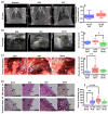
Progression of pleural injury in the RH model. (a) CT images showing baseline non-injured lungs (A) and extensive opacification of the right; ipsilateral hemothorax in which RH was induced; moreover, opacification persists in the RH4, RH7, and RH10 models with limited resolution related to a combination of pleural fluid, atelectasis, or RH. (B,C). A negative change in % indicates a decrease in baseline lung volume at each model endpoint (D). No CT data were available for RH10 due to technological issues precluding imaging. L = Left; R = Right. (b) Representative ultrasonographic images display the retained clot (RC), lung (L), and heart (H) at each endpoint (AC). Morphometric analysis of sonographs from each model was compared (D). (c) Gross presentation of the pleural cavity was examined, showing the retained clot (RC), lung (L), and pleural adhesions (AC, yellow arrows). The pleural adhesion counts included fibrin strands, webs, and sheets which were indicative of pleural organization (D). (d) Naïve and injured rabbit lung tissue were stained with Hematoxylin and Eosin (A1D1, H&E) and Masson Trichome Stain (A2D2, Trichrome) to demonstrate pleural thickening and collagen deposition, respectively. Black arrows indicate the thickening in the pleural lining. Blue-colored stain represents the collagen deposition in the pleural lining (A2D2). All histological images were imaged at 4× objective with a 1 mm scale bar. Morphometric analysis compared the pleural thickening between naïve and RH injured groups (E). Mann–Whitney (panel (d), E) and Kruskal–Wallis with Dunn’s multiple comparison (panel (b), D; panel (d), E; panel (d)) tests determined the statistical significance of the datasets, with *, **, and **** representing p < 0.05, p < 0.01, and p < 0.0001, respectively.
Figure 3
Figure 3
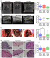
Effects of sex and age on the development of RH at 7 days. Similar to our initial RH7 experiments with young female rabbits (n = 22), RH was also induced in young male (n = 8) and aged female (n = 5) rabbits to determine the contribution of age and sex to RH development. (a) CT images comparing the opacification at right hemithorax between age/sex (AC). Percent changes in the baseline lung volume of rabbits with RH for 7 d were evaluated between age/sex (D). L = Left and R = Right. (b) Ultrasonography at 7 d was performed on all age/ender groups to detect the lung (L), retained clot (RC), and heart (H). 2D Clot area at the 7 d endpoint was measured and compared between the age/sex groups (AD). (c) Gross examination of the lung (L) retained clot (RC) and pleural adhesions (yellow arrows) was evaluated to confirm the ultrasonographic readings (AC). As previously mentioned, the presence of pleural adhesions such as fibrin strands, sheets, and webs was indicative of pleural injury (D). (d) Histology of the injured lung tissue was compared between the age and sex groups. Hematoxylin and Eosin (H&E, A1C1) staining and imaging at 4× demonstrate pleural thickening (black arrows, 1 mm scale). Morphometry demonstrates the differences in pleural thickening between age/sex groups (D). Staining with Masson’s Trichrome (A2C2) suggests overexpression of collagen (blue fibrillar material) associated with peripheral atelectasis of the lung parenchyma. Representative images are illustrated in each panel, as the findings were observed in all rabbits in these groups. Kruskal–Wallis test with Dunn’s multiple comparison test demonstrated statistical difference with * representing p < 0.05.
Figure 4
Figure 4
Cellular analysis and evaluation of the PFs of young and aged rabbits with RH for 7 days. Total red blood cell (RBC) (A) and white blood cell (WBC) (B) counts with differentials (CF) were collected and analyzed from young and aged rabbit PFs with RH for 7 days. Kruskal–Wallis test with Dunn’s multiple comparison test was used to determine statistical significance of the dataset, representing p < 0.05 as * and p < 0.001 as ***.
Figure 5
Figure 5
Analysis and comparison of the biomarkers of inflammation in the PFs of young and aged rabbits with RH for 7 days. The following markers of inflammation were quantified: Total (A) and active (B) PAI-1, TNF-α (C), IL-8 (D), IL-6 (E), TGF-β (F), Glucose (G), LDH activity (H), and total protein (I). Kruskal–Wallis with Dunn’s multiple comparison test determined the statistical significance of the datasets, with *, **, representing p < 0.05 and p < 0.01, respectively.
Figure 6
Figure 6
Comparison of the cellular profiles of RH rabbit models and traumatic hHTX PFs. Red (RBCs, (A)) and White Blood (WBCs, (B)) cell counts and WBC differentials (CF) of rabbit RH models and human hemothorax (hHTX) PFs were evaluated as previously described [30,31]. The lack of PF samples and technological difficulties impeded data collection in RH4, RH7, and RH10 models and clinical subjects. Kruskal–Wallis with Dunn’s multiple comparison tests determined the statistical significance of the datasets, with *, ***, and **** representing p < 0.05, p < 0.001, and p < 0.0001, respectively.
Figure 7
Figure 7
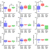
Comparison between the inflammatory and biochemical profile of RH rabbit models and hHTX PFs. Total (A) and active (B) PAI-1, TNF-α (C), IL-8 (D), IL-6 (E), TGF-β (F), Glucose (G), LDH activity (H), and total protein (I) in PFs of rabbit RH models and traumatic hHTX were assayed as previously described [14,30]. Likewise, data point collection in the rabbit RH models and clinical subjects was impeded by the lack or minimal amount of pleural effusion in the model (<1 mL). Prominently, RH4 and RH10 PF samples were unavailable, elucidating the lack of glucose (RH4, n = 2, (G)), LDH activity (RH4, n = 3, (H)), and total protein (RH4, n = 2; RH10, n = 10, (I)) data points. IL-8 data points (RH4, n = 1; RH7, n = 3) that were below the detection level (<1.9 pg/mL) were excluded from analysis (C). Kruskal–Wallis with Dunn’s multiple comparison tests determined the statistical significance of the datasets, with *, **, ***, and **** representing p < 0.05, p < 0.01, p < 0.001, and p < 0.0001, respectively.

References

    1. Rossmann M., Altomare M., Pezzoli I., Abruzzese A., Spota A., Vettorello M., Cioffi S.P.B., Virdis F., Bini R., Chiara O., et al. Risk Factors for Retained Hemothorax after Trauma: A 10-Years Monocentric Experience from First Level Trauma Center in Italy. J. Pers. Med. 2022;12:1570. doi: 10.3390/jpm12101570. - DOI - PMC - PubMed
    1. Zeiler J., Idell S., Norwood S., Cook A. Hemothorax: A Review of the Literature. Clin. Pulm. Med. 2020;27:1–12. doi: 10.1097/CPM.0000000000000343. - DOI - PMC - PubMed
    1. Prakash P.S., Moore S.A., Rezende-Neto J.B., Trpcic S., Dunn J.A., Smoot B., Jenkins D.H., Cardenas T., Mukherjee K., Farnsworth J., et al. Predictors of retained hemothorax in trauma: Results of an Eastern Association for the Surgery of Trauma multi-institutional trial. J. Trauma Acute Care Surg. 2020;89:679–685. doi: 10.1097/TA.0000000000002881. - DOI - PubMed
    1. Choi J., Villarreal J., Andersen W., Min J.G., Touponse G., Wong C., Spain D.A., Forrester J.D. Scoping review of traumatic hemothorax: Evidence and knowledge gaps, from diagnosis to chest tube removal. Surgery. 2021;170:1260–1267. doi: 10.1016/j.surg.2021.03.030. - DOI - PubMed
    1. DuBose J., Inaba K., Okoye O., Demetriades D., Scalea T., O’Connor J., Menaker J., Morales C., Shiflett T., Brown C., et al. Development of posttraumatic empyema in patients with retained hemothorax: Results of a prospective, observational AAST study. J. Trauma Acute. Care Surg. 2012;73:752–757. doi: 10.1097/TA.0b013e31825c1616. - DOI - PubMed

LinkOut - more resources