. 2023 Dec 30;25(1):542.
doi: 10.3390/ijms25010542.

Visualizing the 4D Impact of Gold Nanoparticles on DNA

Affiliations

Visualizing the 4D Impact of Gold Nanoparticles on DNA

Hosam Abdelhady et al. Int J Mol Sci. .

Abstract

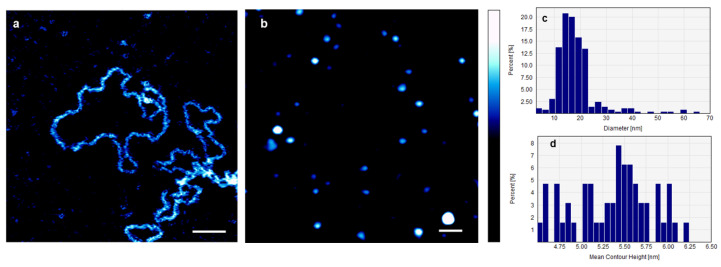
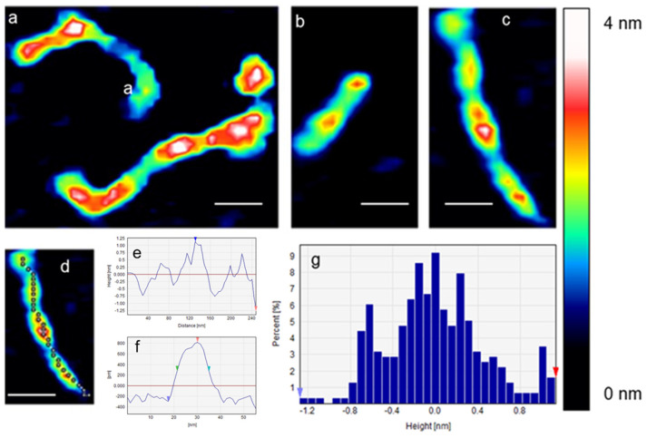
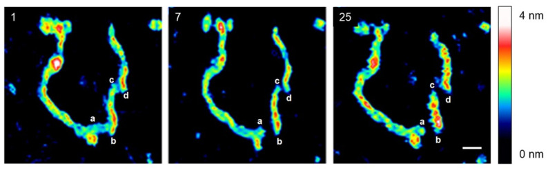
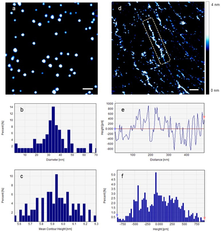
The genotoxicity of AuNPs has sparked a scientific debate, with one perspective attributing it to direct DNA damage and another to oxidative damage through reactive oxygen species (ROS) activation. This controversy poses challenges for the widespread use of AuNPs in biomedical applications. To address this debate, we employed four-dimensional atomic force microscopy (4DAFM) to examine the ability of AuNPs to damage DNA in vitro in the absence of ROS. To further examine whether the size and chemical coupling of these AuNPs are properties that control their toxicity, we exposed individual DNA molecules to three different types of AuNPs: small (average diameter = 10 nm), large (average diameter = 22 nm), and large conjugated (average diameter = 39 nm) AuNPs. We found that all types of AuNPs caused rapid (within minutes) and direct damage to the DNA molecules without the involvement of ROS. This research holds significant promise for advancing nanomedicines in diverse areas like viral therapy (including COVID-19), cancer treatment, and biosensor development for detecting DNA damage or mutations by resolving the ongoing debate regarding the genotoxicity mechanism. Moreover, it actively contributes to the continuous endeavors aimed at fully harnessing the capabilities of AuNPs across diverse biomedical fields, promising transformative healthcare solutions.

Keywords: AFM; AuNPs; COVID-19; Lewis acid; ROS; atomic force microscope; biosensors; cancer; genotoxicity; gold nanoparticles.


Conflict of interest statement

The authors declare no conflicts of interest.

Figures

Figure 1
Figure 1
(a) AFM image of individual DNA plasmids in 0.1× PBS containing 1 mM NiCl2 before the addition of AuNPs. Their average deconvoluted width and height were 4.2 ± 0.7 nm and 2.3 ± 0.5 nm, respectively. (b) AFM image of AuNPs on mica, in 0.1× PBS. The image shows individual uniformly distributed AuNPs. The average diameter of the AuNPs = 16.57 ± 3.74 nm, and their average contour height = 5.4 ± 0.7 nm. Aggregates of AuNPs are also visible in the image. (c,d) Illustration of the distributions of the average diameters and the average contour heights of AuNPs, respectively. Scale bar = 100 nm, and the color bar = 10 nm.
Figure 2
Figure 2
(ac) AFM images of the effect of sAuNPs on individual DNA plasmids in 0.1× PBS. The tracking length of (d) is seen in (e), and the width and height of the cross-section of (d) are seen in (f). The height distribution of (d) is seen in (g). Scale bar = 100 nm.
Figure 3
Figure 3
The time-lapse sequence (1–25 min) of AFM images capturing the effect of AuNPs on an individual DNA plasmid in a 0.1× PBS solution. Initially, after 1 min of adding AuNPs to the DNA solution, beads-on-a-string features were observed, likely due to DNA packaging around the AuNPs. Subsequently, at 7 min, the DNA molecule began to fragment at specific positions (a–b and c–d), possibly indicating the formation of double-strand breaks (DSBs). Over time, the length of the b–c fragment decreased, while its height and width increased, suggesting that the DNA molecule was experiencing stress.
Figure 4
Figure 4
(a) LAuNPs, (b,c) the diameter and contour height of LAuNPs, respectively, (d) the effect of LAuNPs on DNA plasmids imaged on freshly cleaved mica in 0.1× PBS, the dashed rectangle highlights a substantial segment of the damaged DNA molecule, and (e,f) the average contour length and height of the labeled molecule, respectively. Scale bar = 100 nm.
Figure 5
Figure 5
The effect of LCAuNPs on a single DNA plasmid imaged on freshly cleaved mica in 0.1× PBS. (a) LCAuNPs, (b,c) the diameter and contour height of LCAuNPs, respectively, (d) the effect of LCAuNPs on a single DNA plasmid, (eg) are different fragments of the DNA molecule depicted in (d), the dashed rectangle in (f) shows the interaction between a DNA fragment and two AuNPs, and (hj) the average contour length and height of the labeled molecular fragments, respectively. The colored arrowheads in subfigures (hj) correspond to those in subfigures (eg), respectively. Scale bar = 300 nm.

Similar articles

Cited by

References

    1. Chompoosor A., Saha K., Ghosh P.S., MacArthy D.J., Miranda O.R., Zhu Z.J., Arcaro K.F., Rotello V.M. The Role of Surface Functionality on Acute Cytotoxicity, ROS Generation and DNA Damage by Cationic Gold Nanoparticles. Small. 2010;6:2246–2249. doi: 10.1002/smll.201000463. - DOI - PMC - PubMed
    1. Herdt A.R., Drawz S.M., Kang Y., Taton T.A. DNA Dissociation and Degradation at Gold Nanoparticle Surfaces. Colloids Surf. B Biointerfaces. 2006;51:130–139. doi: 10.1016/j.colsurfb.2006.06.006. - DOI - PubMed
    1. Kang B., Mackey M.A., El-Sayed M.A. Nuclear Targeting of Gold Nanoparticles in Cancer Cells Induces DNA Damage, Causing Cytokinesis Arrest and Apoptosis. J. Am. Chem. Soc. 2010;132:1517–1519. doi: 10.1021/ja9102698. - DOI - PubMed
    1. Samhadaneh D.M., Alqarni K.A., Smart A., Kuang M., Moujaber O., Maysinger D., Stochaj U. Gold Nanourchins Induce Cellular Stress, Impair Proteostasis and Damage RNA. Nanomed. Nanotechnol. Biol. Med. 2019;22:102083. doi: 10.1016/j.nano.2019.102083. - DOI - PubMed
    1. Goodman C.M., Chari N.S., Han G., Hong R., Ghosh P., Rotello V.M. DNA-Binding by Functionalized Gold Nanoparticles: Mechanism and Structural Requirements. Chem. Biol. Drug Des. 2006;67:297–304. doi: 10.1111/j.1747-0285.2006.00372.x. - DOI - PubMed