

Long-Term Exposure to Supraphysiological Levels of Testosterone Impacts Rat Submandibular Gland Proteome
- PMID: 38203721
- PMCID: PMC10778877
- DOI: 10.3390/ijms25010550
Long-Term Exposure to Supraphysiological Levels of Testosterone Impacts Rat Submandibular Gland Proteome
Abstract
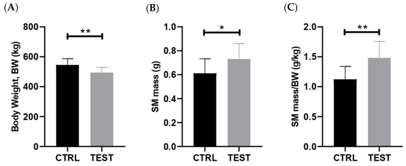
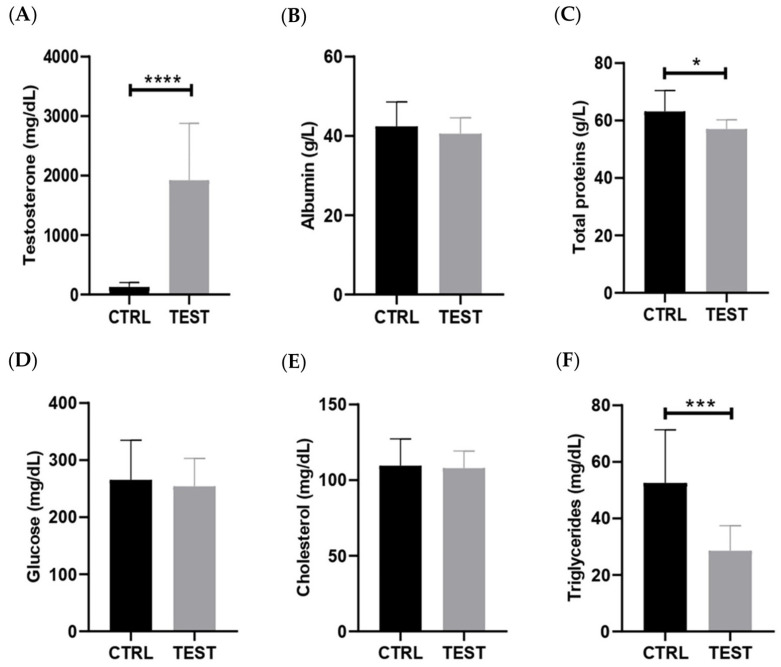
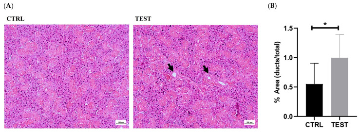
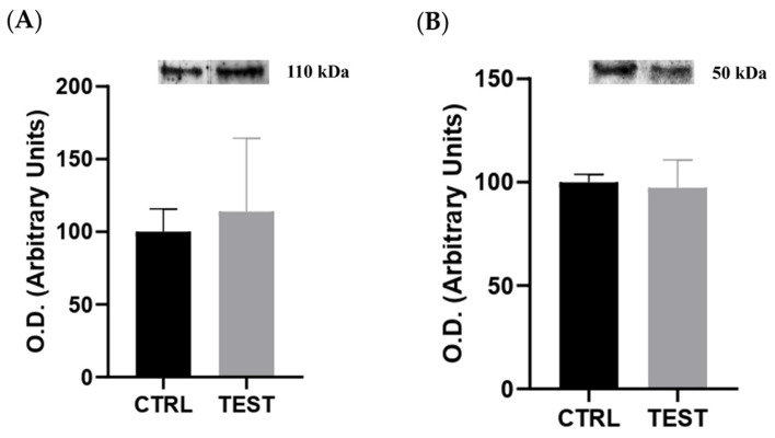
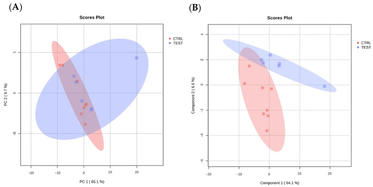
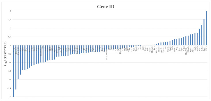
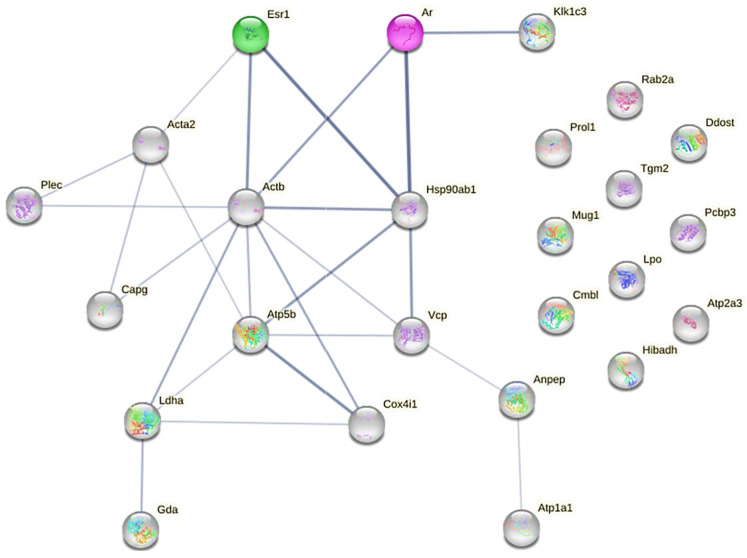
The salivary glands play a central role in the secretion of saliva, whose composition and volume affect oral and overall health. A lesser-explored dimension encompasses the possible changes in salivary gland proteomes in response to fluctuations in sex hormone levels. This study aimed to examine the effects of chronic exposure to testosterone on salivary gland remodeling, particularly focusing on proteomic adaptations. Therefore, male Wistar rats were implanted with subcutaneous testosterone-releasing devices at 14 weeks of age. Their submandibular glands were histologically and molecularly analyzed 47 weeks later. The results underscored a significant increase in gland mass after testosterone exposure, further supported by histologic evidence of granular duct enlargement. Despite increased circulating sex hormones, there was no detectable shift in the tissue levels of estrogen alpha and androgen receptors. GeLC-MS/MS and subsequent bioinformatics identified 308 proteins in the submandibular glands, 12 of which were modulated by testosterone. Of note was the pronounced upregulation of Klk3 and the downregulation of Klk6 and Klk7 after testosterone exposure. Protein-protein interaction analysis with the androgen receptor suggests that Klk3 is a potential target of androgenic signaling, paralleling previous findings in the prostate. This exploratory analysis sheds light on the response of salivary glands to testosterone exposure, providing proteome-level insights into the associated weight and histological changes.
Keywords: GeLC-MS/MS; kallikrein-3; rats; salivary glands; submandibular gland’s proteome; testosterone.
Conflict of interest statement
The authors declare no conflicts of interest.
Figures
Similar articles
-
Effects of a supraphysiological dose of testosterone cypionate on salivary gland function in adult male Wistar rats.J Steroid Biochem Mol Biol. 2024 Oct;243:106587. doi: 10.1016/j.jsbmb.2024.106587. Epub 2024 Jul 14. J Steroid Biochem Mol Biol. 2024. PMID: 39004377
-
The role of cyclic AMP response element-binding protein in testosterone-induced differentiation of granular convoluted tubule cells in the rat submandibular gland.Arch Oral Biol. 2001 Jun;46(6):495-507. doi: 10.1016/s0003-9969(01)00013-9. Arch Oral Biol. 2001. PMID: 11311197
-
DNA Damage and Proteomic Profile Changes in Rat Salivary Glands After Chronic Exposure to Inorganic Mercury.Biol Trace Elem Res. 2022 Sep;200(9):3983-3995. doi: 10.1007/s12011-021-02986-7. Epub 2022 Jan 10. Biol Trace Elem Res. 2022. PMID: 35013890
-
Marked Differences in the Submandibular Salivary Proteome between Sardinian Alcohol-Preferring and Sardinian Alcohol-Non Preferring Rats Revealed by an Integrated Top-Down-Bottom-Up Proteomic Platform.J Proteome Res. 2018 Jan 5;17(1):455-469. doi: 10.1021/acs.jproteome.7b00632. Epub 2017 Nov 16. J Proteome Res. 2018. PMID: 29083190
-
Effect of photic stimuli on rat salivary glands. Role of sympathetic nervous system.Acta Odontol Latinoam. 2000;13(1):3-19. Acta Odontol Latinoam. 2000. PMID: 11885465 Review.
References
MeSH terms
Substances
LinkOut - more resources
Full Text Sources
Miscellaneous
