. 2023 Dec 31;25(1):551.
doi: 10.3390/ijms25010551.

A Plasmid-Borne Gene Cluster Flanked by Two Restriction-Modification Systems Enables an Arctic Strain of Psychrobacter sp. to Decompose SDS

Affiliations

A Plasmid-Borne Gene Cluster Flanked by Two Restriction-Modification Systems Enables an Arctic Strain of Psychrobacter sp. to Decompose SDS

Robert Lasek et al. Int J Mol Sci. .

Abstract

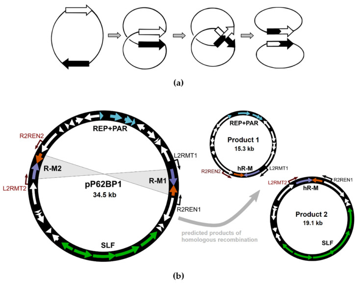
The cold-adapted Psychrobacter sp. strain DAB_AL62B, isolated from ornithogenic deposits on the Arctic island of Spitsbergen, harbors a 34.5 kb plasmid, pP62BP1, which carries a genetic SLF module predicted to enable the host bacterium to metabolize alkyl sulfates including sodium dodecyl sulfate (SDS), a common anionic surfactant. In this work, we experimentally confirmed that the pP62BP1-harboring strain is capable of SDS degradation. The slfCHSL genes were shown to form an operon whose main promoter, PslfC, is negatively regulated by the product of the slfR gene in the absence of potential substrates. We showed that lauryl aldehyde acts as an inducer of the operon. The analysis of the draft genome sequence of the DAB_AL62B strain revealed that the crucial enzyme of the SDS degradation pathway-an alkyl sulfatase-is encoded only within the plasmid. The SLF module is flanked by two restriction-modification systems, which were shown to exhibit the same sequence specificity. We hypothesize that the maintenance of pP62BP1 may be dependent on this unique genetic organization.

Keywords: Psychrobacter; SDS degradation; alkyl sulfatase; psychrophile; restriction–modification system.


Conflict of interest statement

The authors declare no conflict of interest. The funders had no role in the design of the study; in the collection, analyses, or interpretation of data; in the writing of the manuscript; or in the decision to publish the results.

Figures

Figure 1
Figure 1
(a) Genetic organization of plasmid pP62BP1. Arrows indicate the open reading frames. Colors correspond to the main genetic modules of the plasmid: replication and partition module (REP + PAR); restriction–modification systems (R-M1, R-M2); and the SLF module. (b) Predicted metabolic pathway catalyzed by the products of genes identified within the SLF module (slfCHSL) [17]. (c) Growth of Psychrobacter sp. DAB_AL62B and DAB_AL43 (negative control) on LB medium supplemented with SDS to the final concentration of 0.005% (0.35 mM) and Stains-All dye. Zones of SDS degradation are visible as purple areas (image saturation was increased to 130% to emphasize the difference in color).
Figure 2
Figure 2
Growth of Psychrobacter sp. DAB_AL62B (green lines) and DAB_AL43B (negative control; black/grey lines) in the liquid medium supplemented with SDS to the final concentration of 0.01%. The bacterial cultures were preincubated for 4 h at 22 °C. Then, the cultures were divided into two flasks each, and SDS was added to one of the flasks, in which the OD600 was subsequently monitored (solid lines, squares). The flasks without SDS supplementation served as controls (solid lines, triangles). The SDS concentration in supplemented cultures was monitored concurrently (dashed lines, circles). The values plotted are the means of three replicates; error bars represent standard deviation.
Figure 3
Figure 3
(a) Transcriptional organization of the SLF module. The orientation of the predicted promoter sequences is marked by green arrows. The letters A–I indicate binding sites for the primers used in the RT-PCR analysis, including the primers used for a negative control reaction (A + B, in red) and the primer used for cDNA synthesis (C, in blue). (b) Agarose gel electrophoresis of RT-PCR products. The analysis was performed using total RNA isolated from E. coli DH5α (pSLF1) grown in LB supplemented with SDS. The sizes of the amplified fragments were confirmed by comparison with the marker lane. Control reactions were performed with Dnase-treated RNA and pSLF1 DNA templates (negative and positive controls, respectively).
Figure 4
Figure 4
Identification of promoters within the slfCHSL gene cluster and the influence of the expression of the slfR gene in trans on their activity. The graph presents the β-galactosidase activity in E. coli MC1000 carrying (i) the derivatives of the promoter probe vector pRS551 for lacZ transcriptional fusion with the PslfC, PslfH, PslfS, and PslfL promoters, and (ii) the same plasmids in the presence in trans of plasmid pCF-slfR (induction of slfR expression) or pCF-ΔslfR (induction of mutated slfR expression). The values plotted are means of 8 replicates; error bars indicate standard deviation.
Figure 5
Figure 5
Induction of the PslfC promoter by lauryl aldehyde. The graph presents the β-galactosidase activity in E. coli MC1000 carrying the derivative of the promoter probe vector pRS551 for lacZ transcriptional fusion with the PslfC promoter in the presence of the slfR gene in cis at different concentrations of lauryl aldehyde added to the culture medium. The results are expressed as the fraction of the activity observed for E. coli MC1000 (pRS-PslfC). The values plotted are means of 8 replicates; error bars indicate standard deviation.
Figure 6
Figure 6
Digestion of pET-MT1 (a) and pET-MT2 (b) plasmid DNA by pP62BP1-encoded REases. The plasmid DNA was extracted from E. coli BL21(DE3). In both cases, digestion by the ScrFI restriction enzyme and the pP62BP1-encoded REases (RE1, RE2) was observed only in the case of the plasmid DNA isolated after the induction of MTase expression. A number of expected digestion products are visible, including the two largest fragments (2462 bp and 695 bp) indicated by red arrows.
Figure 7
Figure 7
Genetic organization of each of the pP62BP1 R-M systems and the activity of putative promoter sequences in E. coli MC1000 and Psychrobacter sp. DAB_AL43B. The locations of 5′-CCNGG-3′ sites, present in each of the systems, are marked. The range and orientation of the sequences tested for promoter activity are indicated. The values plotted are means of 8 replicates; error bars indicate standard deviation. Values for the MC1000 strain were divided by 10 in order to maintain the same axis.
Figure 8
Figure 8
Hypothetical homologous recombination between the pP62BP1 R-M systems. (a) Theoretical model of DNA recombination between homologous sequences found with the same orientation in a circular DNA molecule. (b) Predicted products of the homologous recombination between the pP62BP1 R-M systems. Hypothetical ‘hybrid’ R-M systems (hR-M) are indicated. Neither of the predicted products was identified experimentally. Binding sites for primers used for PCR-based screening are indicated (cf. Figure S7). Colors correspond to the main genetic modules of the plasmid.

Similar articles

References

    1. Schoch C.L., Ciufo S., Domrachev M., Hotton C.L., Kannan S., Khovanskaya R., Leipe D., Mcveigh R., O’neill K., Robbertse B., et al. NCBI Taxonomy: A comprehensive update on curation, resources and tools. Database J. Biol. Databases Curation. 2020;2020:baaa062. doi: 10.1093/database/baaa062. - DOI - PMC - PubMed
    1. Wang Y., Hou Y., Wang Y., Lu Z., Song C., Xu Y., Wei N., Wang Q. Cloning, expression and enzymatic characteristics of a 2-Cys peroxiredoxin from Antarctic sea-ice bacterium Psychrobacter sp. ANT206. Int. J. Biol. Macromol. 2019;129:1047–1055. doi: 10.1016/j.ijbiomac.2018.09.103. - DOI - PubMed
    1. Linehan M.P., Thoden J.B., Holden H.M. Characterization of two enzymes from Psychrobacter cryohalolentis that are required for the biosynthesis of an unusual diacetamido-d-sugar. J. Biol. Chem. 2021;296:100463. doi: 10.1016/j.jbc.2021.100463. - DOI - PMC - PubMed
    1. Perfumo A., von Sass G.J.F., Nordmann E.L., Budisa N., Wagner D. Discovery and characterization of a new cold-active protease from an extremophilic bacterium via comparative genome analysis and in vitro expression. Front. Microbiol. 2020;11:881. doi: 10.3389/fmicb.2020.00881. - DOI - PMC - PubMed
    1. Tratsiak K., Prudnikova T., Drienovska I., Damborsky J., Brynda J., Pachl P., Kuty M., Chaloupkova R., Rezacova P., Smatanova I.K. Crystal structure of the cold-adapted haloalkane dehalogenase DpcA from Psychrobacter cryohalolentis K5. Pt 5Acta Crystallogr. Sect. F Struct. Biol. Commun. 2019;75:324–331. doi: 10.1107/S2053230X19002796. - DOI - PMC - PubMed

Substances

LinkOut - more resources