. 2023 Dec 31;25(1):552.
doi: 10.3390/ijms25010552.

Exploring the Mechanism of Activation of CFTR by Curcuminoids: An Ensemble Docking Study

Affiliations

Exploring the Mechanism of Activation of CFTR by Curcuminoids: An Ensemble Docking Study

Emanuele Bellacchio. Int J Mol Sci. .

Abstract

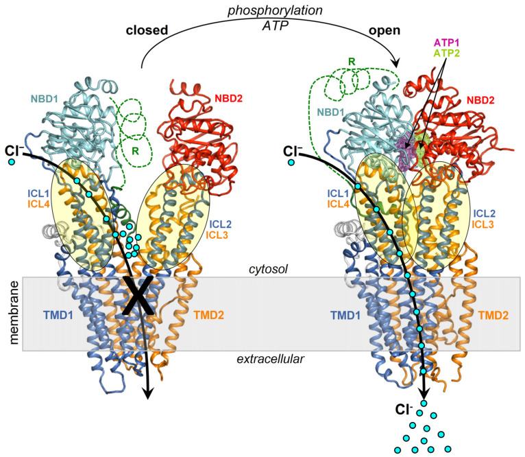
Curcumin, a major constituent of turmeric (Curcuma longa L.), has beneficial effects against several diseases. In cystic fibrosis (CF), this compound improves patients' symptoms by recovering the activity of a number of mutants of the cystic fibrosis transmembrane conductance regulator (CFTR). Despite holding promise in the treatment of CF, the curcumin binding site in CFTR and the molecular mechanism of activation of this channel are still unknown. The results of this study, based on docking and molecular dynamics (MD) simulations, allow us to propose that curcumin binds the closed ATP-free CFTR near the nucleotide-binding domain 1 (NBD1)/ICl1/ICl4 interface. The bound ligand, once approached by the nucleotide-binding domain 2 (NBD2) during transient channel opening, lays at a multiple interdomain cross point. Thereafter, curcumin can bridge NBD1 and NBD2, and also ICL1/ICL4 and ICL2/ICL3, finally tightening the same interdomain interactions that normally uphold the open conformation in the wild-type ATP-bound CFTR. The proposed binding site is compatible with biochemical observations made in previous CFTR-curcumin interaction studies. These findings provide a framework for the design of novel drugs that activate CFTR mutants characterized by defects in ATP binding and/or NBD dimerization or even lacking NBD2.

Keywords: CFTR; CFTR modulators; curcumin; cystic fibrosis; cystic fibrosis transmembrane conductance regulator; docking; molecular dynamics.


Conflict of interest statement

The author declares no conflict of interest.

Figures

Figure 1
Figure 1
Structures of the closed and open CFTR. Shown are the structures of closed CFTR (Protein Data Base, PDB, 5UAK) and open CFTR (PDB 6MSM), highlighting domains and ICLs with a schematic representation of activation. The protein is coloured by domain (TMD1, blue; TMD2, orange; NBD1, cyan; NBD2, red; other regions in grey).
Figure 2
Figure 2
NBD1/ICL1/ICL4 is conformationally invariant in the closed and open CFTR. Closed and open CFTR (respectively, PDB 5UAK and 6MSM, represented as backbone lines with the domains in different colors; the solved R domain fragment and bound ATP molecules are not shown). The conformationally invariant regions (highlighted by cartoons) are defined as those whose Cα atoms overlap within 2.5 Angstroms in the closed and open CFTR protein structures superposed relative to NBD1.
Figure 3
Figure 3
Docking scheme. Curcuminoid structures (diketo curcumin, keto-enol curcumin, and BSc3596) employed in docking (the different chemical moieties in the linkers of the 2-methoxyphenol groups are highlighted by the azure boxes) and CFTR protein targets (ensembles of conformers of the open CFTR, open CFTR-ΔNBD2 construct, and closed CFTR; each ensemble included a parent PDB structure, PDB 6MSM for the open CFTR and the open CFTR-ΔNBD2, and PDB 5UAK for the closed CFTR, the energy minimized and ten MD simulation snapshots of the parent PDB structure; see Section 4). The parent PDB structures are portrayed and colored by the domain (solved R domain fragment and ATP are not shown). The conformationally invariant regions (showing the same conformation in the closed and open CFTR; please see the text) are highlighted by ribbons, and the remainder of the structures by backbone atom lines. The space covered by the docking searches (transparent spheres) encompassed 33.0 Angstroms from the NBD1 center of mass.
Figure 4
Figure 4
Top-scoring curcuminoid docking results on multi-conformer ensembles of the open CFTR, open CFTR-ΔNBD2, and closed CFTR. Shown are the aggregated top-scoring docking results of the individual ligands on each CFTR multi-conformer ensemble (for each of the twelve protein structure targets comprised in a CFTR ensemble, ten top-scoring docking results, out of the ranked 5000 ligand binding poses were kept; subsequently, all groups of ten top-scoring results from each of the twelve docking searches were aggregated). The top-scoring docking results are represented by green spheres (drawn on the centers of docking poses with radii proportional to the normalized docking scores). For simplicity, only the parent PDB protein structures used to derive the CFTR protein ensembles are shown (PDB 6MSM for the open CFTR and open CFTR-ΔNBD2; PDB 5UAK for the closed CFTR; the solved fragment of the R domain is not shown; the two ATP binding sites are highlighted by magenta and dark-yellow meshes in the open CFTR structure).
Figure 5
Figure 5
Identification of a consensus binding pose on the closed and open CFTR ensembles. Shown are the aggregated top binding poses of curcuminoids after applying the criterion that the same binding mode must be found on both the closed and open CFTR conformer ensembles (binding poses are shown as green sticks highlighted by the ovals): (A) diketo curcumin; (B) keto-enol curcumin; (C) BSc3596; (D) binding poses from panels (AC) obeying the additional criterion of binding similarity between curcumin (any of the two tautomers) and BSc3596: only one binding pose fulfills both criteria (and both curcumin tautomers adopts that pose). The displayed protein structures are the open CFTR (PDB 6MSM) and the closed CFTR (PDB 5UAK). The ATP nearest to the consensus curcuminoid binding site is indicated by the magenta meshes in the two bottom protein structures.
Figure 6
Figure 6
Detailed view of the consensus binding pose. The consensus binding pose of a curcuminoid (sticks and green surface) is shown on the closed CFTR (PDB 5UAK) and open CFTR (PDB 6MSM). The protein is coloured by domain (TMD1, blue; TMD2, orange; NBD1, cyan; NBD2, red; other regions in grey). The protein regions with invariant conformation in the closed and open CFTR (please see the text) are represented by ribbons, the other regions as backbone atom lines. The position of the ATP cofactor in the binding site nearest to the curcuminoid consensus binding pose is indicated by the magenta meshes. The NBD2 domain in the rotated views on the right is in front of the viewer and, for clarity, is not shown.
Figure 7
Figure 7
MD simulations of curcuminoids complexed with the closed CFTR. Structures of the three curcuminoids placed in the consensus binding site of the closed CFTR used to kickstart the MD simulations and the same complexes after 100 ns of simulation. The protein is coloured by domain (TMD1, blue; TMD2, orange; NBD1, cyan; NBD2, red). The protein residues near the ligands are shown as sticks (the same color as the parent domains); residues participating in non-bonding interactions (dotted lines) with the ligand are labeled in bold and underscored; other nearby residues requiring only minor movements to engage in interactions with the ligands (as judged by visual inspection) are labelled normally. The ligands are represented as balls and sticks (carbon atoms are in green, oxygens in red, and nitrogens in blue).
Figure 8
Figure 8
MD simulations of curcuminoids complexed with the open CFTR. Structures of the three curcuminoids placed in the consensus binding site of the open CFTR used to kickstart the MD simulations and the same complexes after 100 ns of simulation. For details on protein/residue colors and labels, please refer to the legend in Figure 7.
Figure 9
Figure 9
Energies of interaction and binding free energies. Shown are the interaction energies between the curcuminoids (diketo curcumin, keto-enol curcumin, and BSc3596) and CFTR (decomposed in the individual contributions by the ICL1, ICL4, NBD1, ICL2, ICL3, and NBD2 protein regions) calculated on MD simulation snapshots (the zero-energy value is highlighted by red dashed lines). (A) Interaction energies between curcuminoids and the open CFTR. (B) Interaction energies between curcuminoids and the closed CFTR. The individual protein regions employed in the interaction energy calculations are indicated by an arrow and highlighted with ribbons (the rest of the protein is in backbone lines) in the molecular structure next to each energy plot. The protein is colored by domain (TMD1, blue; TMD2, orange; NBD1, cyan; NBD2, red). The consensus binding site position is indicated by a curcuminoid ligand (L, green meshes). (C) Binding affinities between the curcuminoids and the full CFTR protein calculated on MD snapshots taken every 10 ns of simulation (the time 0 ns corresponds to the energy-minimized structure kickstarting the MD simulations). (D) Calculated binding affinities between the full CFTR (open and closed) and the curcuminoids (averages from panel (C)) or drugs or the two ATP cofactors (as bound in their cryo-EM complexes with CFTR available in the PDB database).

Similar articles

References

    1. Li C., Ramjeesingh M., Wang W., Garami E., Hewryk M., Lee D., Rommens J.M., Galley K., Bear C.E. ATPase activity of the cystic fibrosis transmembrane conductance regulator. J. Biol. Chem. 1996;271:28463–28468. doi: 10.1074/jbc.271.45.28463. - DOI - PubMed
    1. Ostedgaard L.S., Baldursson O., Vermeer D.W., Welsh M.J., Robertson A.D. A functional R domain from cystic fibrosis transmembrane conductance regulator is predominantly unstructured in solution. Proc. Natl. Acad. Sci. USA. 2000;97:5657–5662. doi: 10.1073/pnas.100588797. - DOI - PMC - PubMed
    1. Cheng S.H., Rich D.P., Marshall J., Gregory R.J., Welsh M.J., Smith A.E. Phosphorylation of the R domain by cAMP-dependent protein kinase regulates the CFTR chloride channel. Cell. 1991;66:1027–1036. doi: 10.1016/0092-8674(91)90446-6. - DOI - PubMed
    1. Mathews C.J., Tabcharani J.A., Chang X.B., Jensen T.J., Riordan J.R., Hanrahan J.W. Di-basic protein kinase A sites regulate bursting rate and nucleotide sensitivity of the cystic fibrosis transmembrane conductance regulator chloride channel. J. Physiol. 1998;508:365–377. doi: 10.1111/j.1469-7793.1998.365bq.x. - DOI - PMC - PubMed
    1. Mense M., Vergani P., White D.M., Altberg G., Nairn A.C., Gadsby D.C. In vivo phosphorylation of CFTR promotes formation of a nucleotide-binding domain heterodimer. EMBO J. 2006;25:4728–4739. doi: 10.1038/sj.emboj.7601373. - DOI - PMC - PubMed