. 2023 Dec 31;25(1):558.
doi: 10.3390/ijms25010558.

Ectromelia Virus Affects the Formation and Spatial Organization of Adhesive Structures in Murine Dendritic Cells In Vitro

Affiliations

Ectromelia Virus Affects the Formation and Spatial Organization of Adhesive Structures in Murine Dendritic Cells In Vitro

Zuzanna Biernacka et al. Int J Mol Sci. .

Abstract

Ectromelia virus (ECTV) is a causative agent of mousepox. It provides a suitable model for studying the immunobiology of orthopoxviruses, including their interaction with the host cell cytoskeleton. As professional antigen-presenting cells, dendritic cells (DCs) control the pericellular environment, capture antigens, and present them to T lymphocytes after migration to secondary lymphoid organs. Migration of immature DCs is possible due to the presence of specialized adhesion structures, such as podosomes or focal adhesions (FAs). Since assembly and disassembly of adhesive structures are highly associated with DCs' immunoregulatory and migratory functions, we evaluated how ECTV infection targets podosomes and FAs' organization and formation in natural-host bone marrow-derived DCs (BMDC). We found that ECTV induces a rapid dissolution of podosomes at the early stages of infection, accompanied by the development of larger and wider FAs than in uninfected control cells. At later stages of infection, FAs were predominantly observed in long cellular extensions, formed extensively by infected cells. Dissolution of podosomes in ECTV-infected BMDCs was not associated with maturation and increased 2D cell migration in a wound healing assay; however, accelerated transwell migration of ECTV-infected cells towards supernatants derived from LPS-conditioned BMDCs was observed. We suggest that ECTV-induced changes in the spatial organization of adhesive structures in DCs may alter the adhesiveness/migration of DCs during some conditions, e.g., inflammation.

Keywords: Ectromelia virus; dendritic cells; focal adhesions; migration; podosomes.


Conflict of interest statement

The authors declare no conflicts of interest.

Figures
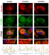
Figure 1
Figure 1
Types of podosomes in BMDCs. The fixed cells were permeabilized and stained for vinculin (red fluorescence), F-actin (green fluorescence), and nuclear DNA (Hoechst 33342, blue fluorescence). The magnified images are of the boxed regions. The fluorescence intensity of vinculin (red line), F-actin (green line), and DNA (blue line) was measured along the yellow line. Scale bars: 10 µm.
Figure 2
Figure 2
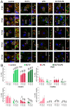
ECTV infection leads to podosome dissolution in BMDCs. Fluorescence microscopy analysis of podosomes and focal adhesions (FAs) in DCs during ECTV replication cycle. (A) Cells uninfected, ECTV-infected, or/and lipopolysaccharide (LPS)-treated were stained for actin (green fluorescence), vinculin (red fluorescence), and DNA (blue fluorescence) at 4, 8, 12, 18, and 24 h post-infection (hpi). The magnified images are of the boxed regions. Arrows—podosomes (white), viral factories (pink; arrowheads—FAs. Scale bars: 10 µm. (B) The mean percentage of cells with no podosomes or having single, clusters, or rosettes podosomes in uninfected or ECTV-infected cells, untreated or treated with LPS. The percentage of cells displaying no podosomes or different types of podosomes was counted in at least 100 random cells per condition per experiment, and an average (with standard deviation (SD)) of three experiments is shown; * p < 0.5, ** p < 0.01, *** p < 0.001.
Figure 3
Figure 3
ECTV infection inhibits the maturation of BMDCs. (A) Representative histograms showing the major histocompatibility complex class II (MHC II), CD80, and CD86 expression on control or ECTV-infected BMDC, untreated or treated with lipopolysaccharide (LPS) at 24 hpi. Numbers represent the MFI value for a given marker. Grey histograms—isotype controls. (B) Graphs show individual data of mean fluorescence intensity (MFI) of MHC II, CD80, and CD86 molecules from four independent experiments. Significant differences were indicated by horizontal bars between two sets of data (* p < 0.05, ** p < 0.01).
Figure 4
Figure 4
ECTV infection induces elongation and widening of focal adhesions (FAs) in BMDCs. (A) Fluorescence microscopy analysis of FAs in uninfected or ECTV-infected cells, untreated or treated with lipopolysaccharide (LPS). The cells were stained for vinculin (green fluorescence) and paxillin (red fluorescence) at 4 and 24 h post-infection (hpi). The magnified images are of the boxed regions. Arrowheads—FAs. Scale bars: 10 µm. (B) The number of FAs/cell and the mean length and width (both in µm) of FAs in uninfected or ECTV-infected cells, untreated or treated with LPS at 4 and 24 hpi. The number of FAs/cell was counted in at least 50 random single cells per condition per experiment from three independent experiments. The length and width of FAs were counted in at least 250 FAs in random cells (all FAs per single random cell) per condition per experiment from three independent experiments. Each point represents individual data, with the bar indicating the mean values of three independent experiments; * p < 0.5, ** p < 0.01, *** p < 0.001. (C). The correlation between length and width of FAs. Each point represents the length vs. width of a single FA of at least 200 FAs per condition per experiment from three independent experiments.
Figure 5
Figure 5
ECTV infection promotes the formation of focal adhesions (FAs) in long cellular extensions of BMDCs. Fluorescence microscopy analysis of dynamics of formation of long cellular extensions in ECTV-infected cells. The cells were stained for F-actin (green fluorescence), vinculin (red fluorescence), and DNA (blue fluorescence) at 4, 8, 12, 18, and 24 h post-infection (hpi). The magnified images are of the boxed regions. Arrows—cellular extensions (white), viral factories (pink); arrowheads—FAs. Scale bars: 10 µm.
Figure 6
Figure 6
Types of long cellular extensions formed by ECTV-infected cells and localization of FAs within them. (A) Thin, (B) tickened end, (C) wide, and (D) branched long extensions. The cells were stained for F-actin, α-tubulin or ECTV antigens (green fluorescence), vinculin, paxillin, mitochondria (Mito) and acetylated-α-tubulin (ac-α-tubulin) (red fluorescence), and DNA (blue fluorescence), or analyzed using scanning electron microscopy (SEM) at 24 h post-infection (hpi). The magnified images are of the boxed regions. Arrows—long cellular extensions (white), viral factories (pink); arrowheads—FAs (white), viral particles (pink). Scale bars: 10 µm.
Figure 7
Figure 7
Types of short cellular extensions formed by ECTV-infected cells and localization of FAs within them. (A) Actin tails and (B) dendrites. The cells were stained for F-actin, α-tubulin or ECTV antigens (green fluorescence), vinculin, paxillin, mitochondria (Mito) and acetylated-α-tubulin (ac-α-tubulin) (red fluorescence), and DNA (blue fluorescence), or analyzed using scanning electron microscopy (SEM) at 24 h post-infection (hpi). The magnified images are of the boxed regions. Arrows—long cellular extensions (white), actin tails (yellow), dendrites (red), viral factories (pink); arrowheads—FAs (white), viral particles (pink). Scale bars: 10 µm.
Figure 8
Figure 8
ECTV infection does not influence the migratory rate of ECTV-infected BMDCs in a wound healing assay. (A). Representative images of BMDC migration into the scrach region (outlined in red) at 0 and 24 h. (B) Graphs show the mean number of cells migrated into the wound with standard deviation (SD) from three independent experiments; ** p < 0.01, n.s.—non-significant. Scale bars: 100 µm. (C) Fluorescence microscopy analysis of the presence of focal adhesions (FAs) in BMDCs migrated into the wound area (outlined in red). The cells were stained for F-actin (green fluorescence), vinculin (red fluorescence), and DNA (blue fluorescence) at 24 h post-infection (hpi). The magnified images are of the boxed regions. Arrowheads—FAs. Scale bars: 50 µm (upper panel) or 10 µm (lower panel).
Figure 9
Figure 9
ECTV infection accelerates the motility of infected BMDCs toward supernatants derived from the LPS-conditioned BMDCs. (A). Representative microscopic images of cells that migrated through the transwell and remained on the underside of the membrane or migrated to the bottom of the lower well in the migration assay (crystal violet stain). Scale bars: 50 µm (upper panel) or 100 µm (lower panel). BMDCs were uninfected or infected with ECTV for 6 h and then subjected to the Transwell migration assay. After incubation for 12 h, migrated cells were stained on the membrane or on the bottom of the lower chamber with crystal violet and photographed. (B) Graphs show the mean number of cells migrated to the lower chamber in a Transwell migration assay with standard deviation (SD) from three independent experiments; ** p < 0.01, n.s.—non-significant.

Similar articles

Cited by

References

    1. Esteban D.J., Buller R.M.L. Ectromelia Virus: The Causative Agent of Mousepox. J. Gen. Virol. 2005;86:2645–2659. doi: 10.1099/vir.0.81090-0. - DOI - PubMed
    1. Alejo A., Saraiva M., Ruiz-Argüello M.B., Viejo-Borbolla A., de Marco M.F., Salguero F.J., Alcami A. A Method for the Generation of Ectromelia Virus (ECTV) Recombinants: In Vivo Analysis of ECTV VCD30 Deletion Mutants. PLoS ONE. 2009;4:e5175. doi: 10.1371/journal.pone.0005175. - DOI - PMC - PubMed
    1. Buller R.M., Potter M., Wallace G.D. Variable Resistance to Ectromelia (Mousepox) Virus among Genera of Mus. Curr. Top. Microbiol. Immunol. 1986;127:319–322. doi: 10.1007/978-3-642-71304-0_38. - DOI - PubMed
    1. Fenner F., Henderson D.A., Arita I., Jezek Z., Ladnyi I.D.L. Smallpox and Its Eradication. World Health Organization; Geneva, Switzerland: 1988.
    1. Ferrari G., Neukamm J., Baalsrud H.T., Breidenstein A.M., Ravinet M., Phillips C., Rühli F., Bouwman A., Schuenemann V.J. Variola Virus Genome Sequenced from an Eighteenth-Century Museum Specimen Supports the Recent Origin of Smallpox. Philos. Trans. R. Soc. B. 2020;375:20190572. doi: 10.1098/rstb.2019.0572. - DOI - PMC - PubMed