. 2024 Jan 1;25(1):562.
doi: 10.3390/ijms25010562.

Expression of RASSF1A, DIRAS3, and AKAP9 Genes in Thyroid Lesions: Implications for Differential Diagnosis and Prognosis of Thyroid Carcinomas

Affiliations

Expression of RASSF1A, DIRAS3, and AKAP9 Genes in Thyroid Lesions: Implications for Differential Diagnosis and Prognosis of Thyroid Carcinomas

Kamila Soboska et al. Int J Mol Sci. .

Abstract

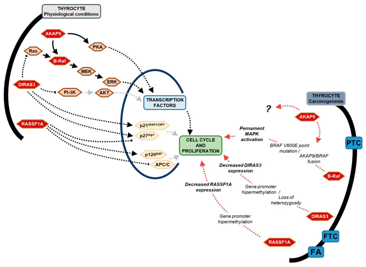
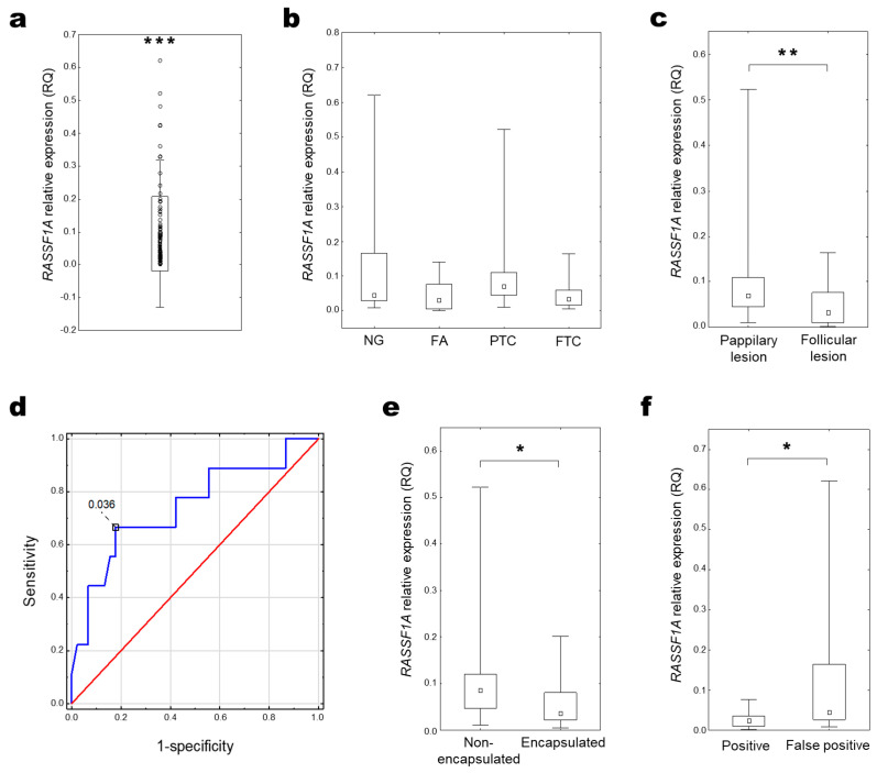
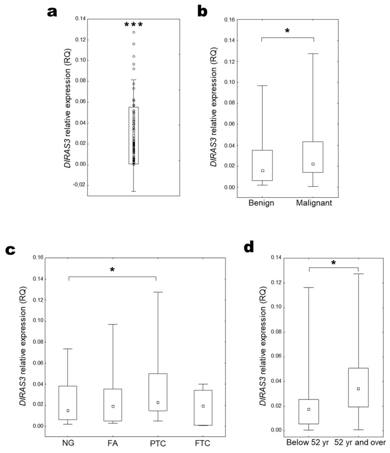
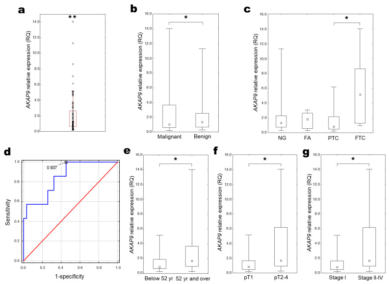
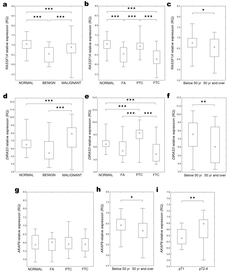
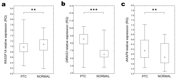
Thyroid carcinoma is the primary endocrine malignancy worldwide. The preoperative examination of thyroid tissue lesion is often unclear. Approximately 25% of thyroid cancers cannot be diagnosed definitively without post-surgery histopathological examination. The assessment of diagnostic and differential markers of thyroid cancers is needed to improve preoperative diagnosis and reduce unnecessary treatments. Here, we assessed the expression of RASSF1A, DIRAS3, and AKAP9 genes, and the presence of BRAF V600E point mutation in benign and malignant thyroid lesions in a Polish cohort (120 patients). We have also performed a comparative analysis of gene expression using data obtained from the Gene Expression Omnibus (GEO) database (307 samples). The expression of RASSF1A and DIRAS3 was decreased, whereas AKAP9's was increased in pathologically changed thyroid compared with normal thyroid tissue, and significantly correlated with e.g., histopathological type of lesion papillary thyroid cancer (PTC) vs follicular thyroid cancer (FTC), patient's age, tumour stage, or its encapsulation. The receiver operating characteristic (ROC) analysis for the more aggressive FTC subtype differential marker suggests value in estimating RASSF1A and AKAP9 expression, with their area under curve (AUC), specificity, and sensitivity at 0.743 (95% CI: 0.548-0.938), 82.2%, and 66.7%; for RASSF1A, and 0.848 (95% CI: 0.698-0.998), 54.8%, and 100%, for AKAP9. Our research gives new insight into the basis of the aggressiveness and progression of thyroid cancers, and provides information on potential differential markers that may improve preoperative diagnosis.

Keywords: AKAP9; BRAF V600E; DIRAS3; RASSF1A; signalling pathways; thyroid carcinoma; thyroid nodules.


Conflict of interest statement

The authors declare no conflict of interest.

Figures

Figure 1
Figure 1
Simplified scheme of physiological action of B-raf kinase, DIRAS3, RASSF1A, and AKAP9 proteins in thyrocyte and their correlation in thyroid carcinogenesis.
Figure 2
Figure 2
RASSF1A expression in thyroid tissue. (a) Distribution of relative RASSF1A expression in all examined samples (RQ) (One sample Wilcoxon test; *** p < 0.001). (b) RASSF1A expression compared among histopathological groups (KW test, p > 0.05) and (c) papillary- and follicular-types of thyroid lesions (UMW test; ** p = 0.01–0.001); (d) ROC curve analysis for RASSF1A expression in differentiation FTC from PTC, where blue line stands for actual ROC curve, and red for diagonal. RASSF1A expression in (e) non-encapsulated and encapsulated tumour groups (UMW test; * p = 0.05–0.01). (f) RASSF1A expression in patients regarding positive and negative (false positive) cytological verification of FNABs (UMW test; * p < 0.05).
Figure 3
Figure 3
DIRAS3 expression in thyroid tissue. (a) Distribution of relative DIRAS3 expression in all examined samples (RQ) (One sample Wilcoxon test; *** p < 0.001). (b) DIRAS3 expression compared among benign and malignant lesions (UMW test, * p = 0.05–0.01) and (c) among histopathological groups (KW test, * p = 0.05–0.01); (d) DIRAS3 expression in patients with malignant lesions depending on the median age (UMW test, * p = 0.05–0.01).
Figure 4
Figure 4
AKAP9 expression in thyroid tissue. (a) Distribution of relative AKAP9 expression in all examined samples (RQ) (One sample Wilcoxon test; ** p = 0.01–0.001). (b) AKAP9 expression compared between benign and malignant lesions (UMW test, * p = 0.05–0.01) and (c) histopathological groups (KW test, * p = 0.05–0.01). (d) ROC curve analysis for AKAP9 expression in differentiation FTC from PTC, where blue line stands for actual ROC curve, and red for diagonal. AKAP9 expression depending on the: (e) median age; (f) tumour size; and (g) tumour stage (UMW test, * p = 0.05–0.01).
Figure 5
Figure 5
RASSF1A (upper row), DIRAS3 (middle row), and AKAP9 (lower row) expression in thyroid tissue (GEO data analysis). Gene expression compared among (a,d) benign (NG and FA) and malignant (PTC and FTC) thyroid changes, (b,e,g) different histopathological types of thyroid tissue lesions (KW test, *** p < 0.001) with unchanged thyroid tissue as the control (NORMAL); Gene expression compared among (c,f,h) groups of patients below (Below 50 y) and over (50 y and over) median age (UMW test, * p = 0.05–0.01; ** p = 0.01–0.001) and (i) cancer stages expressed with the pT scale (UMW test, ** p = 0.01–0.001).
Figure 6
Figure 6
Patient-matched data analysis of gene expression in thyroid tissue (GEO data analysis). (a) RASSF1A, (b) DIRAS3, and (c) AKAP9 expression in thyroid cancer (PTC) compared with patient-matched unchanged thyroid tissue samples (NORMAL) (Wilcoxon matched-pairs signed rank test; ** p = 0.01–0.001; *** p < 0.001).

Similar articles

References

    1. Kebebew E., Weng J., Bauer J., Ranvier G., Clark O.H., Duh Q.Y., Shibru D., Bastian B., Griffin A. The prevalence and prognostic value of BRAF mutation in thyroid cancer. Ann. Surg. 2007;246:466–470. doi: 10.1097/SLA.0b013e318148563d. - DOI - PMC - PubMed
    1. Lee S.T., Kim S.W., Ki C.S., Jang J.H., Shin J.H., Oh Y.L., Kim J.W., Chung J.H. Clinical implication of highly sensitive detection of the BRAF V600E mutation in fine-needle aspirations of thyroid nodules: A comparative analysis of three molecular assays in 4585 consecutive cases in a BRAF V600E mutation-prevalent area. J. Clin. Endocrinol. Metab. 2012;97:2299–2306. doi: 10.1210/jc.2011-3135. - DOI - PubMed
    1. Li M., Dal Maso L., Vaccarella S. Global trends in thyroid cancer incidence and the impact of overdiagnosis. Lancet Diabetes Endocrinol. 2020;8:468–470. doi: 10.1016/S2213-8587(20)30115-7. - DOI - PubMed
    1. Sung H., Ferlay J., Siegel R.L., Laversanne M., Soerjomataram I., Jemal A., Bray F. Global Cancer Statistics 2020: GLOBOCAN Estimates of Incidence and Mortality Worldwide for 36 Cancers in 185 Countries. CA Cancer J. Clin. 2021;71:209–249. doi: 10.3322/caac.21660. - DOI - PubMed
    1. Pizzato M., Li M., Vignat J., Laversanne M., Singh D., La Vecchia C., Vaccarella S. The epidemiological landscape of thyroid cancer worldwide: GLOBOCAN estimates for incidence and mortality rates in 2020. Lancet Diabetes Endocrinol. 2022;10:264–272. doi: 10.1016/S2213-8587(22)00035-3. - DOI - PubMed

MeSH terms

Supplementary concepts