. 2024 Jan 1;25(1):581.
doi: 10.3390/ijms25010581.

Astrocytic Regulation of Endocannabinoid-Dependent Synaptic Plasticity in the Dorsolateral Striatum

Affiliations

Astrocytic Regulation of Endocannabinoid-Dependent Synaptic Plasticity in the Dorsolateral Striatum

Louise Adermark et al. Int J Mol Sci. .

Abstract

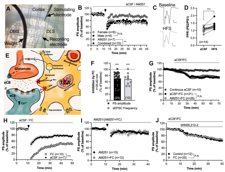
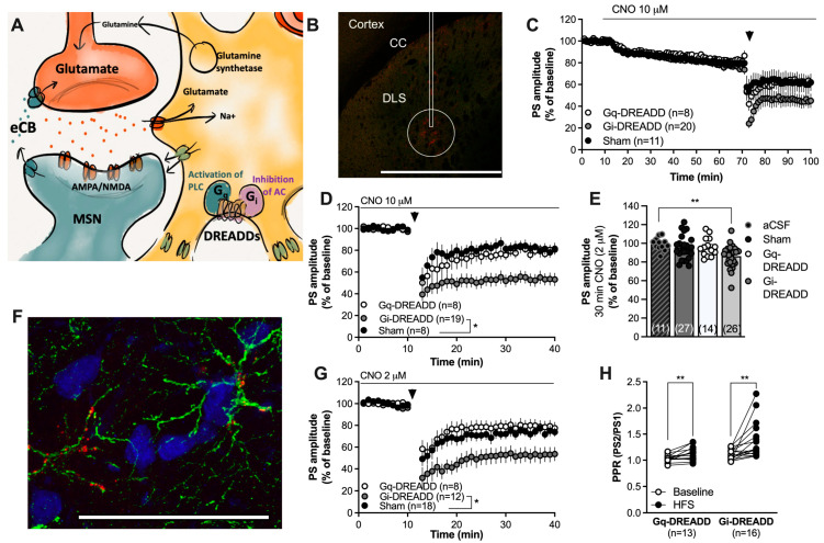
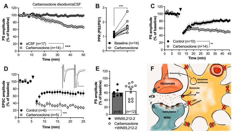
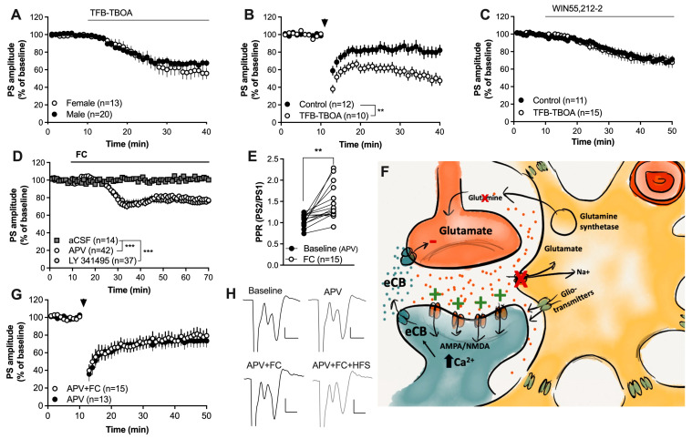
Astrocytes are pivotal for synaptic transmission and may also play a role in the induction and expression of synaptic plasticity, including endocannabinoid-mediated long-term depression (eCB-LTD). In the dorsolateral striatum (DLS), eCB signaling plays a major role in balancing excitation and inhibition and promoting habitual learning. The aim of this study was to outline the role of astrocytes in regulating eCB signaling in the DLS. To this end, we employed electrophysiological slice recordings combined with metabolic, chemogenetic and pharmacological approaches in an attempt to selectively suppress astrocyte function. High-frequency stimulation induced eCB-mediated LTD (HFS-LTD) in brain slices from both male and female rats. The metabolic uncoupler fluorocitrate (FC) reduced the probability of transmitter release and depressed synaptic output in a manner that was independent on cannabinoid 1 receptor (CB1R) activation. Fluorocitrate did not affect the LTD induced by the CB1R agonist WIN55,212-2, but enhanced CB1R-dependent HFS-LTD. Reduced neurotransmission and facilitated HFS-LTD were also observed during chemogenetic manipulation using Gi-coupled DREADDs targeting glial fibrillary acidic protein (GFAP)-expressing cells, during the pharmacological inhibition of connexins using carbenoxolone disodium, or during astrocytic glutamate uptake using TFB-TBOA. While pretreatment with the N-methyl-D-aspartate (NMDA) receptor antagonist 2-amino-5-phosphonopentanoic acid (APV) failed to prevent synaptic depression induced by FC, it blocked the facilitation of HFS-LTD. While the lack of tools to disentangle astrocytes from neurons is a major limitation of this study, our data collectively support a role for astrocytes in modulating basal neurotransmission and eCB-mediated synaptic plasticity.

Keywords: LTD; astrocyte; astroglia; endocannabinoid system; synaptic plasticity.


Conflict of interest statement

Authors declare no conflict of interest.

Figures

Figure 1
Figure 1
Inhibition of the TCA cycle in astrocytes facilitates striatal HFS-LTD. (A) Micrograph demonstrating the position of recording and stimulating electrodes in the DLS. (B) HFS induced LTD in brain slices from both female and male rats in a CB1R-dependent manner. (C) Example traces demonstrating evoked field potentials at baseline (black) and after HFS (gray). Calibration: 0.2 mV, 2 ms. (D) The PPR was significantly enhanced following HFS, indicating that synaptic depression is associated with a decrease in the probability of transmitter release. (E) Schematic and simplified drawing of the tripartite synapse. Striatal astrocytes are extensively interconnected through gap junction channels and form a syncytium that may even include cortical astrocytes. Astrocytes may regulate neurotransmission through the clearance of glutamate by releasing gliotransmitters and by influencing neurovascular coupling. Inhibition of the TCA-cycle in astrocytes using the metabolic uncoupler FC has been demonstrated to decrease the clearance of glutamate resulting in reduced release of the substrate glutamine. Incubation with FC further leads to reduced release of gliotransmitters and bioactive molecules, and impaired astrocytic calcium signaling [39,40,50,51]. (F) Perfusion of FC (5 μM) for 30 min depressed the evoked PS amplitudes and reduced the frequency of glutamatergic inputs onto MSNs. (G) Synaptic depression induced by FC was not CB1R-dependent. (H) Facilitation of HFS-LTD was observed in slices pretreated with FC for at least 60 min and continuously throughout the experiment. (I) The CB1R antagonist prevented HFS-LTD in both aCSF- and FC-treated slices, suggesting that facilitation of LTD is associated with eCB signaling. (J) Synaptic depression induced by the CB1R agonist was not enhanced during the metabolic inhibition of astrocytes. Data are mean values ± sem. Arrows mark time-points for HFS. n = number of recordings, taken from at least four animals. n.s. = not significant. ** p < 0.01; *** p < 0.001.
Figure 2
Figure 2
Chemogenetic activation of Gi-coupled DREADDs in astrocytes facilitates eCB-LTD. (A) Schematic drawing depicting the use of Gq- or Gi-coupled DREADDs to activate pathways that putatively could stimulate or depress astrocytic function. (B) Viral transfection of the DREADD virus was visualized with mCherry (red). The rectangle marks the approximate position of the cannula track; note that there was no damage to the brain tissue following viral injection. While mCherry could be visualized in some cells surrounding the cannula track and even in cells located in the cortex, the circle demonstrates the main area of transfection. Calibration: 1 mm. CC: corpus callosum. (C) Perfusion with CNO (10 μM) slightly depressed the PS amplitude in all treatment groups. (D) Re-baselining-evoked PS amplitudes revealed an enhanced HFS-LTD in CNO-perfused slices expressing Gi-DREADDs targeting GFAP. (E) Bar graph demonstrating the PS amplitudes following bath perfusion with CNO (2 μM) for 30 min. A significant depression of evoked potentials was selectively observed in brain slices expressing Gi-DREADDs. (F) Immunohistochemical staining showing GFAP-expressing astrocytes in green, neurons visualized by NeuN in blue, and the DREADD conjugated with mCherry in red. mCherry was associated with cells expressing GFAP. Calibration: 50 µm. (G) Pretreatment with a lower dose of CNO still facilitated HFS-LTD in slices transfected with Gi-DREADDs targeting GFAP compared to the control. (H) The PPR was enhanced following HFS in both Gq-DREADD- and Gi-DREADD-expressing slices, indicative of the reduced probability for transmitter release. Data are mean values ± sem. Arrows mark time-points for HFS. n = number of recordings, taken from at least four animals. * p < 0.05; ** p < 0.01.
Figure 3
Figure 3
Blockade of connexins facilitates HFS-LTD. (A,B) The gap-junction uncoupler carbenoxolone disodium (100 µM) significantly depressed the evoked PS amplitude and increased the PPR. (C) Pretreatment with carbenoxolone enhanced HFS-LTD. (D) Carbenoxolone-induced facilitation of HFS-LTD at excitatory synapses was confirmed in whole-cell recordings. Note the evoked EPSCs during carbenoxolone perfusion (black) and after HFS (gray) in the upper right panel. Calibration: 100 pA, 25 ms. (E) Carbenoxolone disodium did not facilitate the synaptic depression induced by the CB1R agonist WIN55,212-2 (2 µM). (F) Schematic drawing demonstrating how carbenoxolone treatment would prevent gap-junction coupling and putatively impair the release of gliotransmitters via hemichannels built up by connexins. Data are presented as mean values ± SEM. Arrows mark time-points for HFS. n = number of recordings, taken from at least three animals. ** p < 0.01, *** p < 0.001.
Figure 4
Figure 4
Enhanced glutamatergic signaling may underly the facilitation of HFS-LTD. (A) Inhibition of astrocytic glutamate uptake produced synaptic depression in brain slices from both male and female rats. (B) Slices pretreated with TFB-TBOA were more susceptible to HFS-LTD. (C) TFB-TBOA did not influence the synaptic depression induced by the CB1R agonist. (D) Neither the mGluR2/3 antagonist LY 341495 nor the NMDA receptor antagonist APV prevented the synaptic depression induced by FC. (E) The PPR was significantly enhanced by FC in slices pretreated by FC. (F) Schematic drawing depicting a putative mechanism for how astrocytes may regulate eCB signaling. Inhibition of astrocyte function not only results in impaired glutamate clearance and reduced release of the substrate glutamine, but also the reduced release of gliotransmitters and bioactive molecules, including kynurenic acid, which normally would suppress NMDA receptor activation. Increased levels of glutamate in combination with reduced inhibition of NMDA receptors could lead to increased postsynaptic activation of glutamatergic receptors, resulting in increased postsynaptic calcium, which would promote eCB production and release. (G) APV blocked the ability of FC to promote HFS-LTD. (H) Example traces demonstrating evoked potentials at baseline and following APV (upper traces) and in other slices during incubation in APV + FC followed by HFS (lower traces). Calibration: 0.2 mV, 2 ms. Data are presented as mean values ± SEM. n = number of recordings, taken from at least four animals. Arrows mark time-points for HFS. ** p < 0.01; *** p < 0.001.

Similar articles

Cited by

References

    1. Beard E., Lengacher S., Dias S., Magistretti P.J., Finsterwald C. Astrocytes as Key Regulators of Brain Energy Metabolism: New Therapeutic Perspectives. Front. Physiol. 2021;12:825816. doi: 10.3389/fphys.2021.825816. - DOI - PMC - PubMed
    1. Hansson E., Muyderman H., Leonova J., Allansson L., Sinclair J., Blomstrand F., Thorlin T., Nilsson M., Ronnback L. Astroglia and glutamate in physiology and pathology: Aspects on glutamate transport, glutamate-induced cell swelling and gap-junction communication. Neurochem. Int. 2000;37:317–329. doi: 10.1016/S0197-0186(00)00033-4. - DOI - PubMed
    1. Bergles D.E., Jabs R., Steinhauser C. Neuron-glia synapses in the brain. Brain Res. Rev. 2010;63:130–137. doi: 10.1016/j.brainresrev.2009.12.003. - DOI - PMC - PubMed
    1. Perea G., Yang A., Boyden E.S., Sur M. Optogenetic astrocyte activation modulates response selectivity of visual cortex neurons in vivo. Nat. Commun. 2014;5:3262. doi: 10.1038/ncomms4262. - DOI - PMC - PubMed
    1. Halassa M.M., Fellin T., Takano H., Dong J.H., Haydon P.G. Synaptic islands defined by the territory of a single astrocyte. J. Neurosci. 2007;27:6473–6477. doi: 10.1523/JNEUROSCI.1419-07.2007. - DOI - PMC - PubMed

LinkOut - more resources