

Synthesis of Poly-γ-Glutamic Acid and Its Application in Biomedical Materials
- PMID: 38203869
- PMCID: PMC10779536
- DOI: 10.3390/ma17010015
Synthesis of Poly-γ-Glutamic Acid and Its Application in Biomedical Materials
Abstract
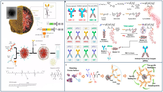
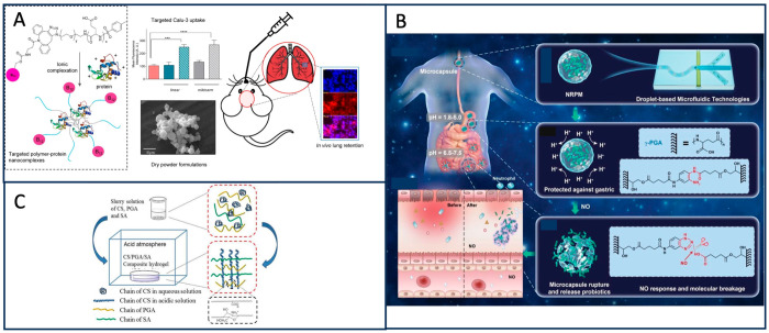
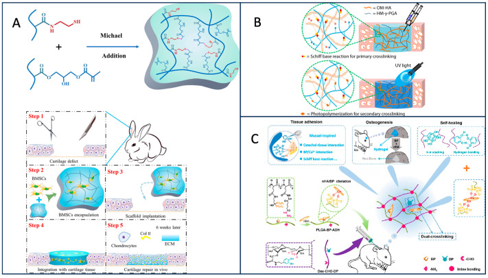
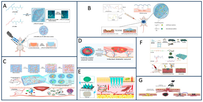
Poly-γ-glutamic acid (γ-PGA) is a natural polymer composed of glutamic acid monomer and it has garnered substantial attention in both the fields of material science and biomedicine. Its remarkable cell compatibility, degradability, and other advantageous characteristics have made it a vital component in the medical field. In this comprehensive review, we delve into the production methods, primary application forms, and medical applications of γ-PGA, drawing from numerous prior studies. Among the four production methods for PGA, microbial fermentation currently stands as the most widely employed. This method has seen various optimization strategies, which we summarize here. From drug delivery systems to tissue engineering and wound healing, γ-PGA's versatility and unique properties have facilitated its successful integration into diverse medical applications, underlining its potential to enhance healthcare outcomes. The objective of this review is to establish a foundational knowledge base for further research in this field.
Keywords: drug delivery; poly-γ-glutamic acid; preparation methods of γ-PGA; tissue engineering; wound healing.
Conflict of interest statement
The authors declare no conflict of interest.
Figures
Similar articles
-
Poly-γ-glutamic acid: production, properties and applications.Microbiology (Reading). 2015 Jan;161(Pt 1):1-17. doi: 10.1099/mic.0.081448-0. Epub 2014 Oct 6. Microbiology (Reading). 2015. PMID: 25288645 Review.
-
Microbial synthesis of poly-γ-glutamic acid: current progress, challenges, and future perspectives.Biotechnol Biofuels. 2016 Jun 29;9:134. doi: 10.1186/s13068-016-0537-7. eCollection 2016. Biotechnol Biofuels. 2016. PMID: 27366207 Free PMC article. Review.
-
Functionalized poly(γ-Glutamic Acid) fibrous scaffolds for tissue engineering.Adv Healthc Mater. 2012 May;1(3):308-15. doi: 10.1002/adhm.201200036. Epub 2012 Apr 5. Adv Healthc Mater. 2012. PMID: 23184745
-
Recent Advances in Microbial Synthesis of Poly-γ-Glutamic Acid: A Review.Foods. 2022 Mar 2;11(5):739. doi: 10.3390/foods11050739. Foods. 2022. PMID: 35267372 Free PMC article. Review.
-
Genetic and metabolic engineering for microbial production of poly-γ-glutamic acid.Biotechnol Adv. 2018 Sep-Oct;36(5):1424-1433. doi: 10.1016/j.biotechadv.2018.05.006. Epub 2018 May 28. Biotechnol Adv. 2018. PMID: 29852203 Review.
Cited by
-
Production and optimization of polyglutamic acid from Bacillus licheniformis: effect of low levels of gamma radiation.AMB Express. 2025 Jun 18;15(1):93. doi: 10.1186/s13568-025-01897-3. AMB Express. 2025. PMID: 40531360 Free PMC article.
-
A Poly-γ-Glutamic Acid/ε-Polylysine Hydrogel: Synthesis, Characterization, and Its Role in Accelerated Wound Healing.Gels. 2025 Mar 22;11(4):226. doi: 10.3390/gels11040226. Gels. 2025. PMID: 40277663 Free PMC article.
-
Poly-γ-Glutamic Acid from a Novel Bacillus subtilis Strain: Strengthening the Skin Barrier and Improving Moisture Retention in Keratinocytes and a Reconstructed Skin Model.Int J Mol Sci. 2025 Jan 24;26(3):983. doi: 10.3390/ijms26030983. Int J Mol Sci. 2025. PMID: 39940752 Free PMC article.
-
Brown Algae as a Valuable Substrate for the Cost-Effective Production of Poly-γ-Glutamic Acid for Applications in Cream Formulations.Polymers (Basel). 2024 Jul 22;16(14):2091. doi: 10.3390/polym16142091. Polymers (Basel). 2024. PMID: 39065408 Free PMC article.
-
Poly (γ) glutamic acid: a unique microbial biopolymer with diverse commercial applicability.Front Microbiol. 2024 Feb 13;15:1348411. doi: 10.3389/fmicb.2024.1348411. eCollection 2024. Front Microbiol. 2024. PMID: 38414762 Free PMC article. Review.
References
-
- Tanaka T., Taniguchi M. Poly(γ-glutamic acid) from Bacillus subtilis as an optically heterogeneous peptide in which D- and L-glutamic acid isomers are copolymerized into a single chain. Hydrocolloids. 2000;33138:459–463.
-
- Ivanovics G., Erdos L. Ein Beitrag zum Wesen der Kaspelsubstanz des Milzbrandbazillus. Z. Immuntatsforsch. 1937;90:5–19.
-
- Ho G.-H., Ho T.-I., Hsieh K.-H., Su Y.-C., Lin P.-Y., Yang J., Yang K.-H., Yang S.-C. γ-Polyglutamic acid produced by Bacillus subtilis (natto): Structural characteristics, chemical properties and biological functionalities. J. Chin. Chem. Soc. 2006;53:1363–1384. doi: 10.1002/jccs.200600182. - DOI
-
- Park C., Choi Y.-H., Shin H.-J., Poo H., Song J.J., Kim C.-J., Sung M.-H. Effect of high-molecular-weight poly-γ-glutamic acid from Bacillus subtilis (chungkookjang) on Ca solubility and intestinal absorption. J. Microbiol. Biotechnol. 2005;15:855–858.
Publication types
LinkOut - more resources
Full Text Sources
