

Acute performance of stylet driven leads for left bundle branch area pacing: A comparison with lumenless leads
- PMID: 38204462
- PMCID: PMC10774671
- DOI: 10.1016/j.hroo.2023.11.014
Acute performance of stylet driven leads for left bundle branch area pacing: A comparison with lumenless leads
Abstract
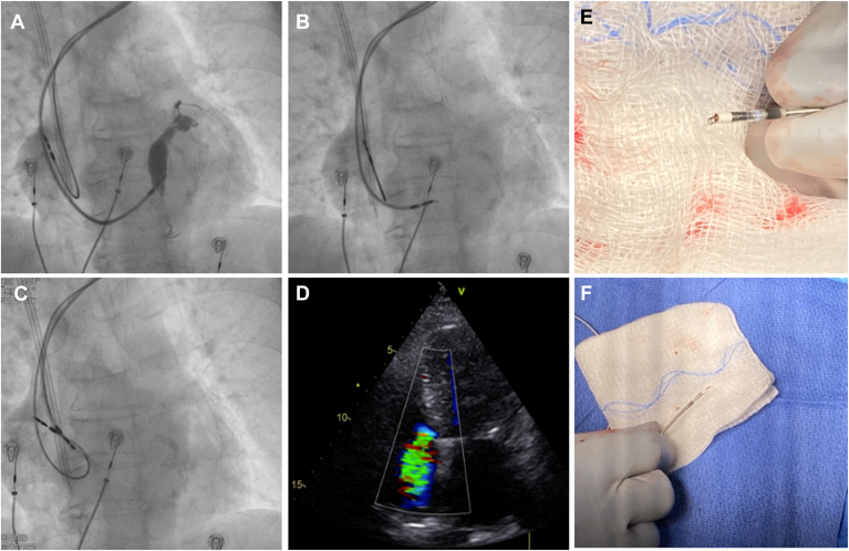
Background: Lumenless leads (LLLs) are widely used for left bundle branch area pacing (LBBAP). Recently, stylet-driven leads (SDLs) have also been used for LBBAP.
Objective: The purpose of this study was to evaluate the acute performance of SDLs during LBBAP in comparison with LLLs.
Methods: Consecutive patients undergoing LBBAP for bradycardia or cardiac resynchronization therapy indications at 2 high-volume, early conduction system pacing adopters, tertiary centers were included from January 2019 to July 2023. Patients received either SDLs or LLLs at the discretion of the implanting physician. Acute performance and follow-up data of both lead types were evaluated.
Results: A total of 925 LBBAP implants were included, 655 using LLLs and 270 using SDLs. Overall, LBBAP acute success was significantly higher with LLLs than SDLs (95.3% vs 85.1%, respectively; P <.001) even after the learning curve (97% vs 86%; P = .013). LLLs were implanted in more mid-basal septal positions in comparison with SDLs, which tended to be implanted in more inferior and mid-apical septal positions. Acute lead-related complications were higher with SDLs than LLLs (15.9% vs 6.1%, respectively; P <.001) with 15 cases of lead damage during implant (4.4% vs 0.5%; P <.001) but decreased with acquired experience and were comparable in the last 100 patients included in each group. Lead implant and fluoroscopy times were shorter for SDLs, with lead dislodgment occurring in 0.9% with LLLs and 1.5% with SDLs (P = .489).
Conclusion: Acute lead performance proved to be different between LLLs and SDLs. A specific learning curve should be considered for SDLs even for implanters with extensive previous experience with LLLs.
Keywords: Conduction system pacing; Left bundle branch area pacing; Lumenless leads; Physiological pacing; Stylet-driven leads.
© 2023 Heart Rhythm Society. Published by Elsevier Inc.
Figures


Similar articles
-
Stylet-Driven Lead Vs. Lumenless Lead for Left Bundle Branch Area Pacing: Systematic Literature Review and Meta-Analysis.Pacing Clin Electrophysiol. 2025 Jul;48(7):682-690. doi: 10.1111/pace.15209. Epub 2025 May 23. Pacing Clin Electrophysiol. 2025. PMID: 40407354
-
Stylet-driven leads versus lumenless pacing leads in patients with left bundle branch area pacing: A systematic review and meta-analysis.Heart Rhythm O2. 2024 Nov 17;6(2):166-175. doi: 10.1016/j.hroo.2024.11.006. eCollection 2025 Feb. Heart Rhythm O2. 2024. PMID: 40231099 Free PMC article.
-
Lead Integrity and Failure Evaluation in Left Bundle Branch Area Pacing: The LIFE-LBBAP Study.JACC Clin Electrophysiol. 2025 Jan;11(1):158-170. doi: 10.1016/j.jacep.2024.09.020. Epub 2024 Nov 20. JACC Clin Electrophysiol. 2025. PMID: 39570266
-
Procedural outcome and follow-up of stylet-driven leads compared with lumenless leads for left bundle branch area pacing.Europace. 2023 Oct 5;25(10):euad295. doi: 10.1093/europace/euad295. Europace. 2023. PMID: 37766468 Free PMC article.
-
Stylet-driven leads compared with lumenless leads for left bundle branch area pacing: a systematic review and meta-analysis.BMC Cardiovasc Disord. 2024 Oct 26;24(1):598. doi: 10.1186/s12872-024-04273-4. BMC Cardiovasc Disord. 2024. PMID: 39462327 Free PMC article.
Cited by
-
Riding the Highs and Lows of the Conduction System Pacing Wave-Our Experience.J Cardiovasc Dev Dis. 2025 Apr 22;12(5):164. doi: 10.3390/jcdd12050164. J Cardiovasc Dev Dis. 2025. PMID: 40422935 Free PMC article.
-
Stylet-driven Leads or Lumenless Leads for Conduction System Pacing.Arrhythm Electrophysiol Rev. 2024 Sep 13;13:e14. doi: 10.15420/aer.2024.18. eCollection 2024. Arrhythm Electrophysiol Rev. 2024. PMID: 39385772 Free PMC article. Review.
-
Conduction system pacing in heart failure: Time for a paradigm shift?Heart Fail Rev. 2025 Mar;30(2):365-380. doi: 10.1007/s10741-024-10469-9. Epub 2024 Nov 23. Heart Fail Rev. 2025. PMID: 39579301 Review.
-
Lumenless and Stylet-Driven Leads for Left Bundle Branch Area Pacing: Materials, Techniques, Benefits, and Trade-Offs of the Two Approaches.J Clin Med. 2024 Aug 13;13(16):4758. doi: 10.3390/jcm13164758. J Clin Med. 2024. PMID: 39200900 Free PMC article. Review.
-
Successful pacemaker implantation using left bundle branch area pacing in a patient with dextrocardia: A case report.J Arrhythm. 2025 Jun 19;41(3):e70118. doi: 10.1002/joa3.70118. eCollection 2025 Jun. J Arrhythm. 2025. PMID: 40538501 Free PMC article.
References
-
- Vijayaraman P., Chelu M.G., Curila K., et al. Cardiac conduction system pacing: a comprehensive update. JACC Clin Electrophysiol. 2023 S2405-500X(23)00391-2. - PubMed
-
- Abdelrahman M., Subzposh F.A., Beer D., et al. Clinical outcomes of His bundle pacing compared to right ventricular pacing. J Am Coll Cardiol. 2018;71:2319–2330. - PubMed
-
- Sharma P.S., Patel N.R., Ravi V., et al. Clinical outcomes of left bundle branch area pacing compared to right ventricular pacing: results from the Geisinger-Rush Conduction System Pacing Registry. Heart Rhythm. 2022;19:3–11. - PubMed
LinkOut - more resources
Full Text Sources
