Review
. 2023 Jul 28:15:143-148.
doi: 10.1016/j.ibneur.2023.07.003. eCollection 2023 Dec.

Where do you measure the Bregma for rodent stereotaxic surgery?

Affiliations
Review

Where do you measure the Bregma for rodent stereotaxic surgery?

Marianna Nogueira Cecyn et al. IBRO Neurosci Rep. .

Abstract

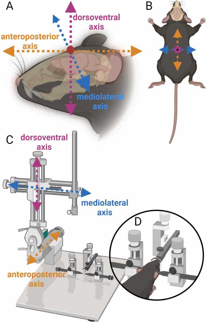
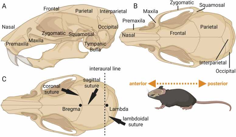
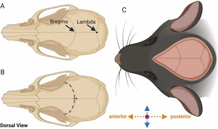
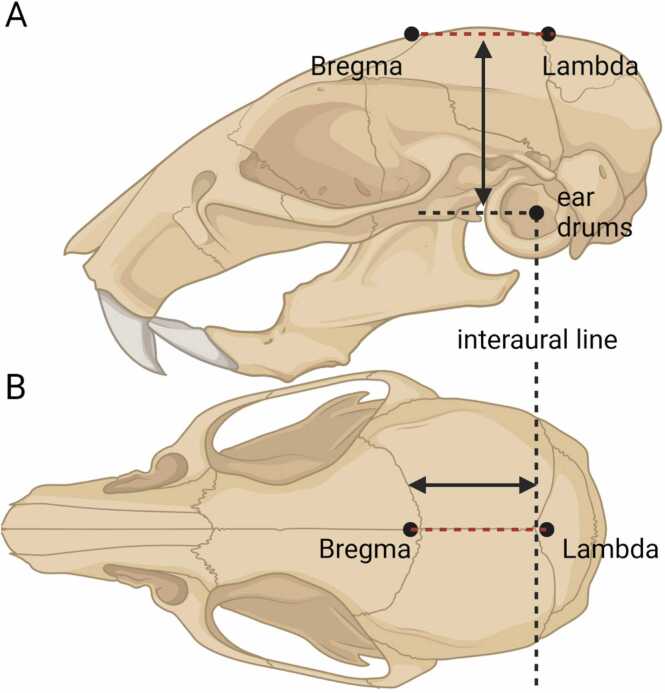
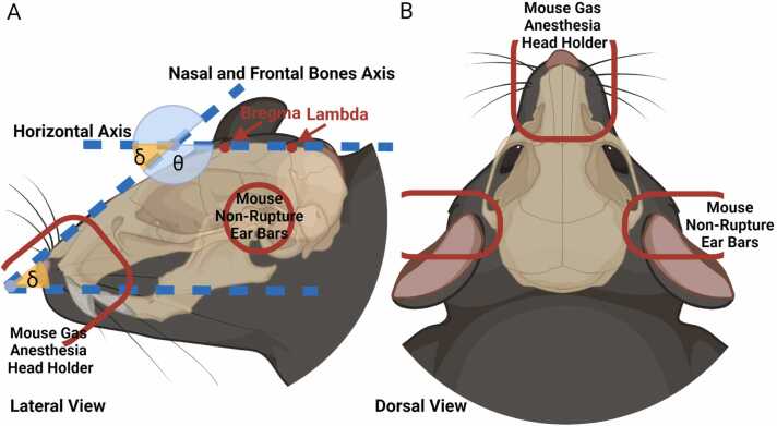
The advent of the stereotaxic apparatus developed by Clarke and Horsley revolutionized neuroscience research, enabling precise 3D navigation along the skull mediolateral, anteroposterior, and dorsoventral axes. In rodents, the Bregma is widely used as the origin reference point for the stereotaxic coordinates, but the specific procedure for its measurement varies among different laboratories. Notably, the renowned brain atlas developed by Paxinos and Franklin lacks explicit instructions on the Bregma determination. Recent studies have found discrepancies in skull and brain landmark measurements. This review describes the commonly used brain atlases and highlights the limitations in accurately measuring the stereotaxic coordinates. In addition, we propose alternative and more reliable approaches to measure the Bregma. It is imperative to address the misconceptions about the accuracy of stereotaxic surgeries, as it can significantly impact a substantial portion of neuroscience research.

Keywords: Animal brain surgery; Brain atlas; Horsley-Clarke’s apparatus; Mouse; Rat; Skull landmarks.


Conflict of interest statement

The authors declare no conflicts of interest.

Figures

Fig. 1
Fig. 1
Mouse skull-body orientation and a rodent stereotaxic apparatus. (A) Lateral view of the mouse head, illustrating its orientation in three axes in relation to the skull and brain. (B) Dorsal view of the mouse body, highlighting the three axes. (C) Illustration of the rodent animal stereotaxic apparatus. (D) Detailed view of the mouse head holder and ears bars. Note: All figures in this review employ a consistent color pattern for the three axes: orange represents the anteroposterior axis, pink represents the dorsoventral axis, and blue represents the mediolateral axis.
Fig. 2
Fig. 2
Anatomical representation of the mouse skull from different perspectives (based on Cook, 1965) (A) Lateral view of the mouse skull, highlighting the bones. (B) Dorsal view of the mouse skull, highlighting the bones. (C) Dorsal view of the rodent skull sutures and landmarks relevant for stereotaxic surgery. Created using BioRender.
Fig. 3
Fig. 3
Dorsal view of the rodent skull diagram with sutures and detailed points of measurement of the Bregma and the Lambda. Bregma and Lambda based on Paxinos and Franklin (2019). Red dot: the Bregma measured on the crossing point between the coronal and the sagittal sutures. Blue dot: the Bregma measured on the crossing pointing between the sagittal suture with the best-fitting parabola on the coronal suture. Based on Blasiak et al. (2010)(C) Skull landmarks on surgery view, it is important to note that during the surgery the research will partially see the skull depending on the incision size. Created using BioRender.
Fig. 4
Fig. 4
Craniometric and stereotaxic skull landmarks and measures. According to MBSC, the Bregma is 3.8 ( ± 0.25) mm rostral and 5.8 ( ± 0.48) from the interaural line, and the lambda is 0.41 ( ± 0.26) mm caudal and 5.8 ( ± 0.48) from the interaural line. (A) Lateral view of mouse skull, Bregma and Lambda dorsoventral distances from the intraural line. (B) Dorsal view of mouse skull, Bregma and Lambda anteroposterior distances from the intraural line.
Fig. 5
Fig. 5
Mouse head position on stereotaxic apparatus. (A) Lateral view of the mouse head. Note that the Bregma and the Lambda are on the same horizontal plane. Nasal and frontal bones are in angle relative to the horizontal plane (δ). The δ and θ are complementary to 180 degrees. The mouse head holder must be in the correct angle with the horizontal plane to coorectly position the mouse head. In red, the approximate position of mouse head holder and ear bars. (B) Dorsal view of the mouse head. In red, the approximate position of mouse head holder and ear bars . Created using BioRender.

Similar articles

Cited by

References

    1. Al-Rodhan N.R.F., Kelly P.J. Pioneers of stereotactic neurosurgery. Stereo Funct. Neurosurg. 1992;58:60–66. doi: 10.1159/000098974. - DOI - PubMed
    1. Arefev R.A., Kiroy V.N., Bulat N.V., Petrushan M.V., Burbelov M.O., Sazhin S.L., Vladimirskiy B.M., Matukhno A.E., Chechevatova V.V., Semynina V.G., Lysenko L.V. Methods for calculating the stereotaxic coordinates of rat brain structures by pixel coordinates of the image obtained by confocal and two-photon laser scanning microscopy. J. Neurosci. Methods. 2021:361. doi: 10.1016/j.jneumeth.2021.109273. - DOI - PubMed
    1. Blasiak T., Czubak W., Ignaciak A., Lewandowski M.H. A new approach to detection of the bregma point on the rat skull. J. Neurosci. Methods. 2010;185:199–203. doi: 10.1016/j.jneumeth.2009.09.022. - DOI - PubMed
    1. Catani M. A little man of some importance. Brain. 2017;140:3055–3061. doi: 10.1093/brain/awx270. - DOI - PMC - PubMed
    1. Chon U., Vanselow D.J., Cheng K.C., Kim Y. Enhanced and unified anatomical labeling for a common mouse brain atlas. Nat. Commun. 2019;10:5067. doi: 10.1038/s41467-019-13057-w. - DOI - PMC - PubMed

LinkOut - more resources