. 2023 Jul 14:15:116-125.
doi: 10.1016/j.ibneur.2023.07.001. eCollection 2023 Dec.

Unprecedented effect of vitamin D3 on T-cell receptor beta subunit and alpha7 nicotinic acetylcholine receptor expression in a 3-nitropropionic acid induced mouse model of Huntington's disease

Affiliations

Unprecedented effect of vitamin D3 on T-cell receptor beta subunit and alpha7 nicotinic acetylcholine receptor expression in a 3-nitropropionic acid induced mouse model of Huntington's disease

Skv Manjari et al. IBRO Neurosci Rep. .

Abstract

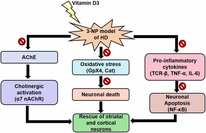
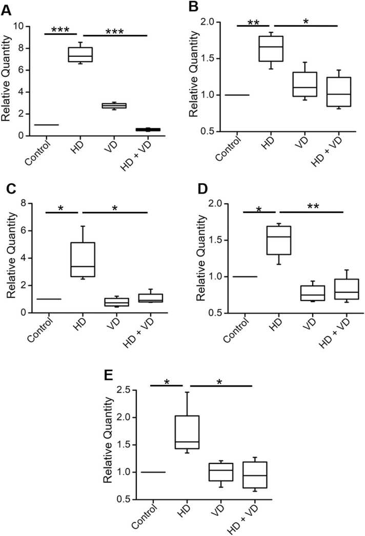
Introduction: 3-NP induction in rodent models has been shown to induce selective neurodegeneration in the striatum followed by the cortex (Brouillet, 2014). However, it remains unclear whether, under such a neurotoxic condition, characterized by neuroinflammation and oxidative stress, the gene expression of the immune resident protein, T-cell receptor beta subunit (TCR-β), α7 nicotinic acetylcholine receptor (α7 nAChRs), the nuclear factor kappa B (NF-κB), inflammatory cytokines (TNF-α and IL-6), and antioxidants (Cat and GpX4) get modulated on Vitamin D3 (VD) supplementation in the central nervous system.

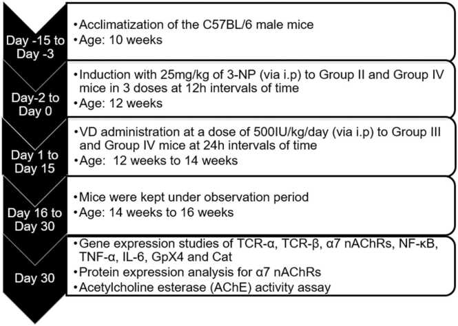
Methods: In the present study, real-time polymerase chain reaction (RT-PCR) was performed to study the expression of respective genes. Male C57BL/6 mice (8-12 weeks) were divided into four groups namely, Group I: Control (saline); Group II: 3-NP induction via i.p (HD); Group III: Vitamin D3 (VD) and Group IV: (HD + VD) (Manjari et al., 2022).

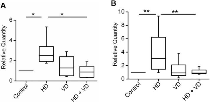
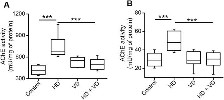
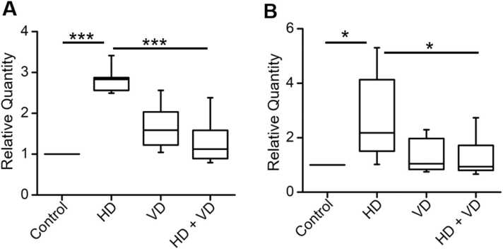
Results: On administration of 500IU/kg/day of VD, HD mice showed a significant reduction in the gene expression of the immune receptor, TCR-β subunit, nuclear factor kappa B (NF-κB), inflammatory cytokines, and key antioxidants, followed by a decrease in the acetylcholinesterase activity.

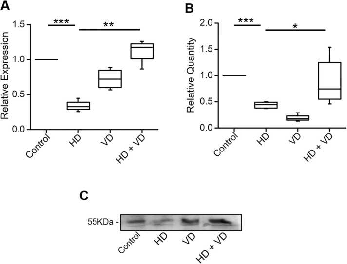
Conclusion: A novel neuroprotective effect of VD in HD is demonstrated by combating the immune receptor, TCR-β gene expression, antioxidant markers, and inflammatory cytokines. In addition, HD mice on VD administration for 0-15 days showed an enhancement in cholinergic signaling with restoration in α7 nAChRs mRNA and protein expression in the striatum and cortex.

Keywords: Huntington’s disease (HD); Immune receptors; Interleukin-6 (IL-6); Nuclear factor kappa B (NF-κB); T-cell receptor-beta subunit (TCR- β); Tumor necrosis factor-alpha (TNF-α); Vitamin D3 (VD); α7 nicotinic acetylcholine receptors (α7 nAChRs).


Conflict of interest statement

The authors declare no conflict of interest.

Figures

ga1
Graphical abstract
Fig. 1
Fig. 1
Timeline and design for the study. C57BL/6 male mice at the age of ten to twelve weeks were undertaken in the present study. Mice were separated into four different groups. Group II and Group IV mice were injected (i.p) with 3-nitropropionic acid (3-NP) at 25 mg/kg dose at 12 h intervals of time (cumulative dose of 75 mg/kg; Manjari et al., 2022). Vitamin D3 (VD; 500IU/kg/day) was supplemented in Group III mice (VD only) and after post-injection of 3-NP to Group IV mice (HD + VD) for 15 days i.e. from Day 1 – Day 15. Mice were kept under observation from Day 1 to Day 30. On the 30th day, mice were sacrificed and the cortical and striatal brain tissue samples were extracted for gene and protein expression analysis.
Fig. 2
Fig. 2
Vitamin D3 (VD) intake decreases the gene expression of the TCR-β subunit in the cortex and striatum of HD mice. (A) Data demonstrating a significant increase in the cortical gene expression of the TCR-β subunit in Group II mice (HD vs control; n = 6, p = 0.009, paired sample t-test). VD administration to Group IV mice post-3-NP injection rescued the mRNA expression of the TCR-β subunit in the cortex of HD mice (HD + VD vs HD; n = 6, p = 0.02, paired sample t-test). (B) RT-PCR results depicting VD administration decreased the mRNA expression of the TCR-β subunit in the striatum of 3-NP induced HD mice (HD + VD vs HD; n = 10, p = 0.008, paired sample t-test). TCR-β subunit expression was significantly upregulated in Group II mice as compared to Group I mice (HD vs control; n = 10, p = 0.005, paired sample t-test). Data is represented as box-and-whisker plots depicting the median with first and third quartiles and whiskers representing the 5th and 95th percentile values.
Fig. 3
Fig. 3
Effect of VD supplementation on the protein and gene expression of α7 nicotinic acetylcholine receptors (α7 nAChRs) in the cortex and striatum of HD mice (A) On the 30th day, an overall change in the protein expression of α7 nAChRs was observed in cortical tissue samples from all the four groups of mice (n = 4, p < 0.001, one-way ANOVA). VD supplementation rescued the cortical expression of α7 nAChRs in Group IV mice (HD + VD) as compared to Group II (HD) mice (n = 4, p < 0.001, paired sample t-test). (B) A significant increase in the mRNA expression of α7 nAChRs was also observed in the striatal samples of 3-NP induced HD mice on VD administration (Group IV vs Group II, n = 6, p = 0.02, paired sample t-test). The mRNA expression of α7 nAChRs got significantly decreased in HD mice when compared with control mice (Group II vs Group I; n = 6, p < 0.001, paired sample t-test). Data is represented as box-and-whisker plots depicting the median in the first and third quartiles and whiskers represent the 5th and 95th percentile values. (C) Representative gel image for protein expression of α7 nAChRs from the cortical tissues.
Fig. 4
Fig. 4
Effect of VD supplementation on the enzymatic activity of acetylcholine esterase (AChE) in the cortex and striatum of HD mice (A) On the 30th day, a notable change in the activity of AChE was observed in the cortex of all four groups of mice (n = 6, p < 0.001, one-way ANOVA). VD induction significantly combated the activity of AChE in Group IV mice as compared to Group II mice (HD + VD vs HD; n = 6, p = 0.002, paired sample t-test). (B) A significant decrease in the activity of AChE was also observed in the striatal brain tissue samples of Group IV mice, supplemented with VD (HD + VD vs HD, n = 8, p < 0.001, paired sample t-test). Data is represented as box-and-whisker plots depicting the median with first and third quartiles and whiskers representing the 5th and 95th percentile values.
Fig. 5
Fig. 5
VD administration rescues the gene expression of antioxidants in HD mice (A) mRNA expression of glutathione peroxidase 4 (GpX4) was increased in Group II mice (HD vs control, n = 6, p < 0.001, paired sample t-test), which subsided on VD supplementation (HD + VD vs HD, n = 6, p < 0.001, paired sample t-test) (B) The mRNA expression of catalase (Cat) was also found to be increased in the cortex of Group II mice as compared to Group I mice (HD vs control, n = 8, p = 0.008, paired sample t-test). The gene expression of Cat got alleviated on VD supplementation in Group IV mice reflecting its antioxidant effect (HD + VD vs HD, n = 8, p = 0.003, paired sample t-test). Data is represented as box-and-whisker plots depicting the median with first and third quartiles and whiskers representing the 5th and 95th percentile values.
Fig. 6
Fig. 6
An Anti-inflammatory effect of 500IU/kg of VD in the striatum and cortex of 3-NP induced HD mice. (A) mRNA expression of nuclear factor kappa B (NF-κB) was significantly increased in Group II mice (HD vs control, n = 4, p < 0.001, paired sample t-test) which got combated on VD supplementation (HD + VD vs HD, n = 4, p < 0.001, paired sample t-test). (B) Group IV mice administered with VD showed a significant reduction in the striatal gene expression of TNF-α (HD + VD vs HD, n = 4, p = 0.02, paired sample t-test) which got elevated in Group II mice injected with 75 mg/kg of 3-NP (HD vs control, n = 4, p = 0.005, paired sample t-test). (C) An increased mRNA expression of another inflammatory cytokine, interleukin 6 (IL-6) was observed in the striatum of Group II mice (HD vs control, n = 4, p = 0.02, paired sample t-test) which got subsided in Group IV mice when administered with VD (HD + VD vs HD, n = 4, p = 0.01, paired sample t-test). (D) A similar increase in the mRNA expression of tumor necrosis factor-α (TNF-α) was observed in the cortical brain tissue samples of Group II mice injected with 3-NP (HD vs control, n = 4, p = 0.01, paired sample t-test). The post-supplementation of VD for 15 days significantly attenuated the gene expression of TNF-α (HD + VD vs HD, n = 4, p = 0.002, paired sample t-test) and (E) interleukin 6 (IL-6) in the cortex of Group II mice (HD + VD vs HD, n = 4, p = 0.01, paired sample t-test). IL-6 gene expression was observed to be highly elevated in Group II mice on 3-NP injection (HD vs control, n = 4, p = 0.03, paired sample t-test). Data is represented as box-and-whisker plots depicting the median with first and third quartiles and whiskers representing 5th and 95th percentile values.

Similar articles

Cited by

References

    1. Ahuja M., Chopra K., Bishnoi M. Inflammatory and neurochemical changes associated with 3-Nitropropionic acid neurotoxicity. Toxicol. Mech. Methods. 2008;18:335–339. doi: 10.1080/15376510701563738. - DOI - PubMed
    1. AlJohri R., AlOkail M., Haq S.H. Neuroprotective role of vitamin D in primary neuronal cortical culture. eNeurologicalSci. 2019;14:43–48. doi: 10.1016/j.ensci.2018.12.004. - DOI - PMC - PubMed
    1. Bakhtiari-Dovvombaygi H., Izadi S., Zare M., Asgari Hassanlouei E., Dinpanah H., Ahmadi-Soleimani S.M., Beheshti F. Vitamin D3 administration prevents memory deficit and alteration of biochemical parameters induced by unpredictable chronic mild stress in rats. Sci. Rep. 2021;11 doi: 10.1038/s41598-021-95850-6. - DOI - PMC - PubMed
    1. Baudouin S.J., Angibaud J., Loussouarn G., Bonnamain V., Matsuura A., Kinebuchi M., Naveilhan P., Boudin H. The signaling adaptor protein CD3zeta is a negative regulator of dendrite development in young neurons. Mol. Biol. Cell. 2008;19:2444–2456. doi: 10.1091/mbc.e07-09-0947. - DOI - PMC - PubMed
    1. Brouillet E. The 3-NP model of striatal neurodegeneration. Curr. Protoc. Neurosci. 2014;67(9):1–14. doi: 10.1002/0471142301.ns0948s67. - DOI - PubMed

LinkOut - more resources