

Computed tomography evaluation of alterations in the masticator space due to invasion by malignant head and neck neoplasms
- PMID: 38204895
- PMCID: PMC10775806
- DOI: 10.1590/0100-3984.2023.0024-en
Computed tomography evaluation of alterations in the masticator space due to invasion by malignant head and neck neoplasms
Abstract
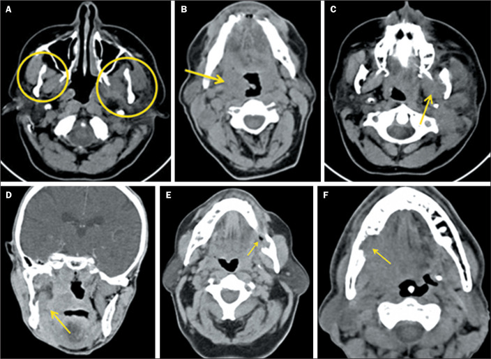
Objective: To evaluate alterations in the masticator space due to the dissemination of malignant neoplasms originating from the tonsillar fossa, retromolar trigone, maxillary sinus, or nasopharynx, using computed tomography (CT), as well as to correlate the presence of trismus with the CT findings and the dimensions of the tumor.
Materials and methods: We evaluated the medical records of 65 patients with malignant tumors in the regions described. The images were analyzed by two physician examiners, working independently, who were blinded to the clinical data. In the evaluation of the masticator space, the following parameters were considered: symmetry with the contralateral space; obliteration of the fat plane, retromolar trigone, or pharyngeal space; edema/atrophy of the medial or lateral pterygoid muscles; and destruction of the mandibular ramus.
Results: Obliteration of the fat plane was found in 69.2% of the patients. Asymmetry, edema/atrophy, and bone destruction were detected in 27.7%, 26.2%, and 20.0% of the patients, respectively. Trismus was identified in 15.4% of the patients. Of the patients with trismus, 90.0% had stage T4 tumors, compared with only 43.8% of those without trismus. Trismus was 11.6 times more common among the patients with stage T4 tumors than among those with lower-stage tumors. Neoplasms of the tonsillar fossa and retromolar trigone collectively accounted for 95.0% of the cases. The CT scans showed edema/atrophy of the pterygoid muscles in 60.0% of the patients with trismus and in 21.8% of those without. An association was observed between T4 tumor stage and edema/atrophy of the pterygoid muscles. In addition, the risk of trismus was 5.4 times higher among the patients with stage T4 tumors.
Conclusion: In our patient sample, the most common finding was obliteration of the fat plane, followed by asymmetry and edema/atrophy. Most of the patients with T4 tumors had trismus, together with edema/atrophy of the pterygoid muscles.
Objetivo: Avaliar, por meio de tomografia computadorizada, alterações do espaço mastigador (EM) decorrentes de disseminação de neoplasias malignas originárias da loja tonsilar, trígono retromolar, seio maxilar e nasofaringe, e correlacionar presença de trismo com achados tomográficos e dimensões do tumor.
Materiais e métodos: Foram selecionados prontuários de 65 pacientes portadores de tumores malignos nas regiões descritas. A análise das imagens foi realizada por dois examinadores médicos, separadamente, sem o conhecimento das informações clínicas. Na avaliação do EM, foram considerados: simetria com o EM contralateral; obliteração do plano gorduroso, do trígono retromolar e do espaço faríngeo; edema e/ou atrofia dos músculos pterigóideos medial e lateral; destruição do ramo da mandíbula.
Resultados: Foram observados obliteração do plano gorduroso em 69,2% dos pacientes, assimetria em 27,7%, espessamento/atrofia em 26,2% e destruição óssea em 20,0%. Presença de trismo foi encontrada em 15,4% dos pacientes. Na associação entre dimensão do tumor e trismo, foram observados trismo em 90,0% dos tumores em estágio T4, enquanto a porcentagem de tumores em estágio T4 sem trismo foi de 43,8%. Pacientes com tumores T4 apresentaram 11,6 vezes mais trismo que os dos demais estágios. Neoplasias da loja tonsilar e trígono retromolar perfizeram 95,0% dos casos. Em 60,0% dos pacientes com trismo havia edema e/ou atrofia dos músculos pterigóideos na tomografia computadorizada e em 21,8% nos sem trismo. Observou-se associação entre tumores T4 e edema e/ou atrofia dos músculos pterigóideos e 5,4 vezes mais chance de apresentarem trismo.
Conclusão: A maioria dos pacientes apresentou obliteração do plano gorduroso, seguido de assimetria e espessamento/atrofia. O trismo estava presente na maioria dos pacientes T4 com espessamento/atrofia dos músculos pterigóideos.
Keywords: Masticatory muscles, Neoplasms; Tomography; Trismus.; X-ray computed, Neoplasms.
Figures
References
-
- Seltzer SE, Wang AM. Modern imaging of the masseter muscle: normal anatomy and pathosis on CT and MRI. Oral Surg Oral Med Oral Pathol. 1987;63:622–629. - PubMed
-
- O’Leary MR. Trismus: modern pathophysiological correlates. Am J Emerg Med. 1990;8:220–227. - PubMed
-
- Ichimura K, Tanaka T. Trismus in patients with malignant tumours in the head and neck. J Laryngol Otol. 1993;107:1017–1020. - PubMed
-
- Wei Y, Xiao J, Zou L. Masticator space: CT and MRI of secondary tumor spread. AJR Am J Roentgenol. 2007;189:488–497. - PubMed
LinkOut - more resources
Full Text Sources