. 2023 Dec 26;12(12):2520-2537.
doi: 10.21037/tlcr-23-707. Epub 2023 Dec 14.

Exosomal ITGB6 from dormant lung adenocarcinoma cells activates cancer-associated fibroblasts by KLF10 positive feedback loop and the TGF-β pathway

Affiliations

Exosomal ITGB6 from dormant lung adenocarcinoma cells activates cancer-associated fibroblasts by KLF10 positive feedback loop and the TGF-β pathway

Xiang Feng et al. Transl Lung Cancer Res. .

Abstract

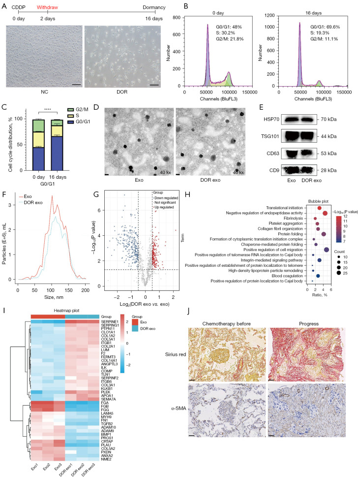
Background: Dormant cancer cells are commonly known to play a pivotal role in cancer recurrence and metastasis. However, the mechanism of tumor dormancy and recurrence remains largely unknown. This study aimed to investigate the mechanism by which exosomes derived from dormant lung adenocarcinoma (LUAD) cells activate cancer-associated fibroblasts (CAFs) to reconstruct the extracellular matrix (ECM), providing a novel idea for decoding the mechanism of tumor dormancy.

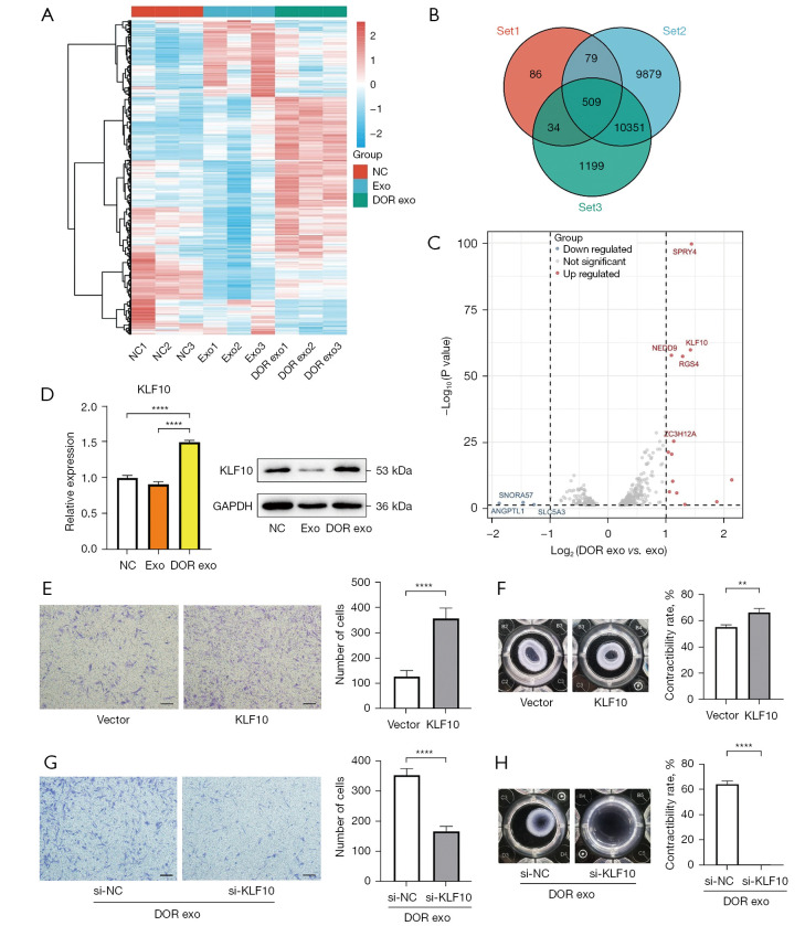
Methods: In this study, high-dose cisplatin was used to induce the dormant LUAD cells. Exosomes were extracted from the culture supernatant of normal and dormant cancer cells. The effects of selected exosomal proteins on the fibroblasts were evaluated. RNA-seq for fibroblasts and exosomal proteomics for normal and dormant cancer cells were used to identify and verify the mechanism of activating fibroblasts.

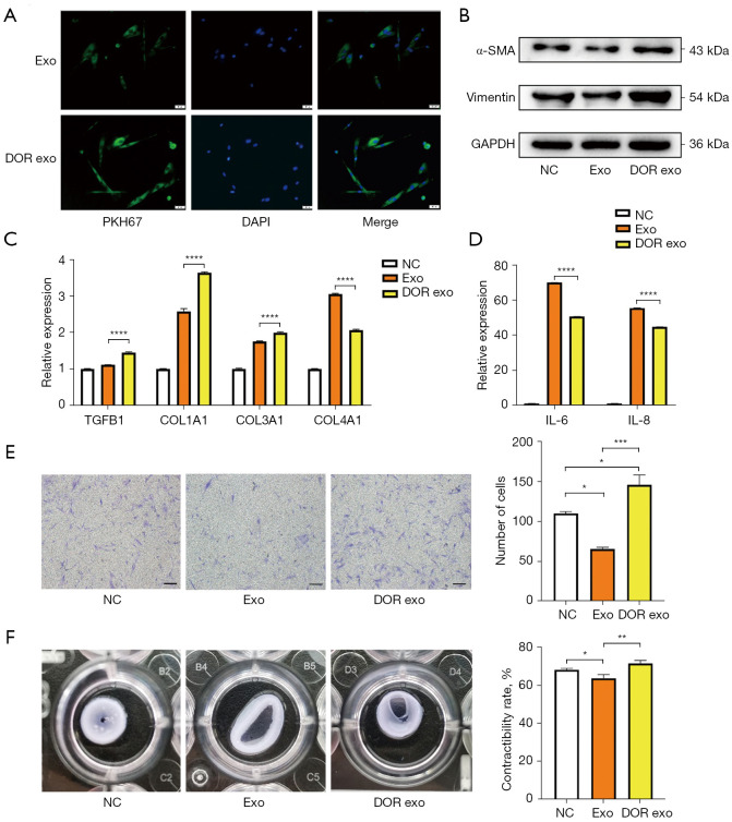
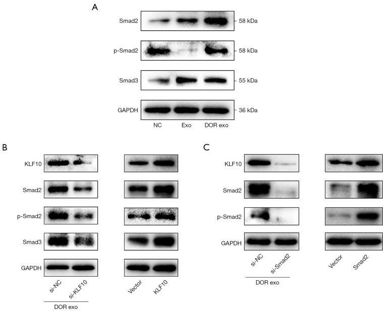
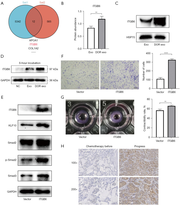
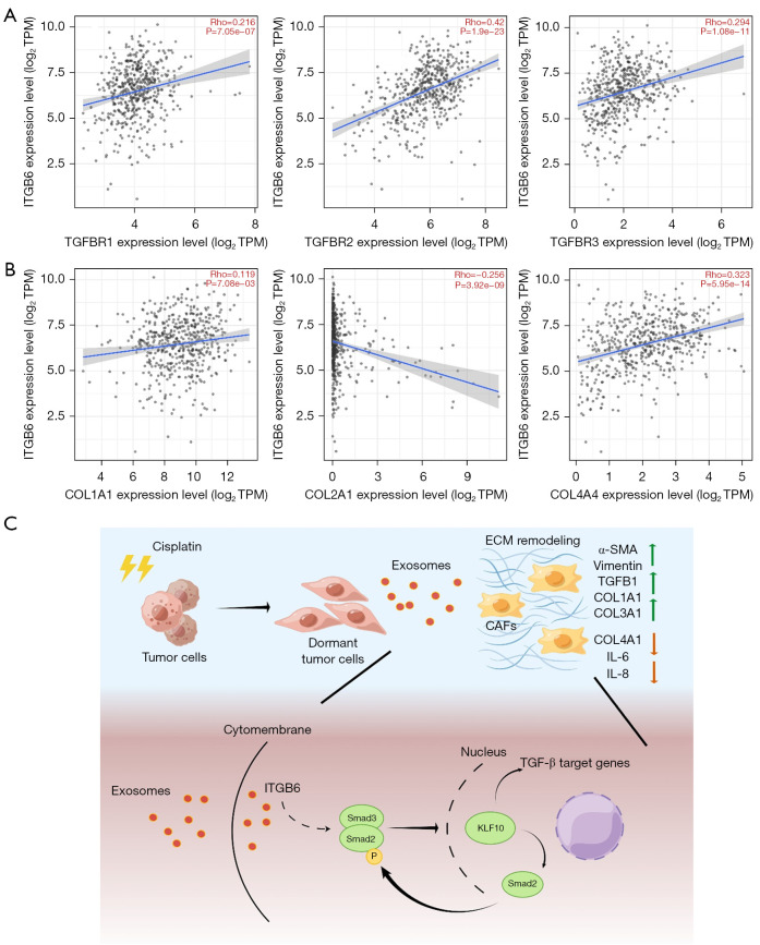
Results: We demonstrated that exosomes derived from dormant A549 cells could be taken by fibroblasts. Exosomal ITGB6 transferred into fibroblasts induced the activation of CAFs by activating the KLF10 positive feedback loop and transforming growth factor β (TGF-β) pathway. High ITGB6 expression was associated with activation of the TGF-β pathway and ECM remodeling.

Conclusions: In all, we demonstrated that CAFs were activated by exosomes from dormant lung cancer cells and reconstruct ECM. ITGB6 may be a critical molecule for activating the TGF-β pathway and remodeling ECM.

Keywords: Lung cancer; cancer-associated fibroblasts (CAFs); dormancy; exosomes.


Conflict of interest statement

Conflicts of Interest: All authors have completed the ICMJE uniform disclosure form (available at https://tlcr.amegroups.com/article/view/10.21037/tlcr-23-707/coif). The authors have no conflicts of interest to declare.

Figures

Figure 1
Figure 1
Exosomes derived from dormant LUAD cells reconstruct the ECM. A549 was treated with a high dose of cis-platinum for 2 days and cultured with fresh complete medium for 16 days. The treatment protocol is shown. (A) Cell morphology is shown in the pictures by inverted light microscope. Magnification: ×20; scale bar =400 µm. (B,C) Cell cycle assay suggested that the S phase population was reduced and cells were arrested in the G0/G1 phase. (D) exo and DORexo were isolated and identified. The morphology of exosomes was observed by transmission electron microscopy. (E) Exosome-associated markers (HSP70, TSG101, CD63, and CD9) were detected by western blot. (F) Nanoparticle tracking analysis showed that the diameters of exosomes secreted from A549 cells and dormant A549 cells range from 50 to 170 nm. (G) The volcano plot shows differentially expressed proteins between exosomes derived from A549 cells and derived from dormant A549 cells (DORexo vs. exo). (H) GO functional enrichment analysis for differential expressed proteins from proteomics was performed and shown as a bubble map. (I) ECM-related proteins from GO enriched dataset were clustered and visualized as a heatmap. (J) Immunohistochemical analysis using the antibody against α-SMA evaluated the infiltration degree of α-SMA positive CAFs of one case of LUAD before treatment or with no chemotherapy response. Sirius red staining was performed to evaluate the degree of ECM remodeling. Magnification: ×100; scale bar =50 µm. ****, P<0.0001. CDDP, cisplatin; NC, A549 cells; DOR, dormant A549 cells; exo, A549-secreted exosomes; DORexo, dormant A549-secreted exosomes; α-SMA, alpha smooth muscle actin; LUAD, lung adenocarcinoma; ECM, extracellular matrix; GO, Gene Ontology; CAFs, cancer-associated fibroblasts.
Figure 2
Figure 2
Exosomes derived from dormant LUAD cells induce the activation of CAFs. MRC-5 was selected as the research object and incubated with exosomes. (A) Immunofluorescence showed that exosomes marked with PKH67 were absorbed by MRC-5. Magnification: ×200; scale bar =50 µm. (B) MRC-5 was incubated with exosomes for 48 hours. Western blotting showed that the CAF markers (α-SMA and Vimentin) were upregulated after treatment with DORexo. (C) Matrix-related molecules in exosome-treated fibroblasts were measured by qRT-PCR. (D) Inflammation-related molecules in exosome-treated fibroblasts were measured by qRT-PCR. (E) The ability of invasion was evaluated by transwell assays. MRC-5 cells were treated with exosomes. Cells that invaded to the bottom surface were stained with crystal violet and observed by microscopy. Magnification: ×40; scale bar =200 µm. The numbers of invading cells were counted from 6 fields of view in each group. (F) The contractile ability of fibroblasts was assessed by ECM-remodeling assay. MRC-5 cells incubated with exosomes were embedded in a matrix mixture for 48 hours. Gel contraction was accessed by taking pictures. The formula for gel contraction value was 100 × (well diameter-gel diameter)/well diameter. Data were presented as the mean ± SD, and analyzed with one-way ANOVA. *, P<0.05; **, P<0.01; ***, P<0.001; ****, P<0.0001. exo, A549-secreted exosomes; DORexo, dormant A549-secreted exosomes; DAPI, 4',6-diamidino-2-phenylindole; NC, negative control; α-SMA, alpha smooth muscle actin; GAPDH, glyceraldehyde-3-phosphate dehydrogenase; LUAD, lung adenocarcinoma; CAFs, cancer-associated fibroblasts; qRT-PCR, quantitative real-time polymerase chain reaction; ECM, extracellular matrix; SD, standard deviation; ANOVA, analysis of variance; IL, interleukin.
Figure 3
Figure 3
KLF10 is the key molecule for activation of CAFs. (A) The RNA expression profile of MRC-5 incubated with exosomes was examined. The heatmap of differentially expressed molecules was presented. (B) Venn diagram showing that the overlapping part between the 3 datasets contained 509 molecules, including our dataset and two independent datasets (GSE116679 and GSE61797). (C) The volcano plot for 509 molecules showed that KLF10 was one of the most representative differentially expressed RNA molecules. (D) The expression of KLF10 was measured by qRT-PCR and western blotting. (E,G) The invasion ability of fibroblast was accessed by transwell assays with crystal violet staining. Magnification: ×40; scale bar =200 µm. (F,H) The contractile ability of fibroblast was accessed by ECM-remodeling assay. Data are presented as the mean ± SD, and analyzed with Student’s t-test. **, P<0.01; ****, P<0.0001. NC, negative control; exo, A549-secreted exosomes; DORexo, dormant A549-secreted exosomes; CAFs, cancer-associated fibroblasts; qRT-PCR, quantitative real-time polymerase chain reaction; ECM, extracellular matrix; SD, standard deviation.
Figure 4
Figure 4
KLF10-Smad2 positive feedback loop induces the activation of CAFs by activating the TGF-β pathway. (A) Protein expression of the TGF-β pathway were measured by western blot analysis. MRC-5 cells were treated with DORexo compared to exo. GAPDH was used as the internal control. (B) Protein expression of KLF10 and TGF-β pathway were measured by western blot analysis. MRC-5 cells incubated with DORexo were transfected with si-KLF10. Further, MRC-5 cells were transfected with KLF10 overexpression plasmid. GAPDH was used as the internal control. (C) Protein expression of KLF10 and TGF-β pathway were measured by western blot analysis. MRC-5 cells incubated with DORexo were transfected with si-Smad2. Further, MRC-5 cells were transfected with Smad2 overexpression plasmid. GAPDH was used as the internal control. NC, negative control; exo, A549-secreted exosomes; DORexo, dormant A549-secreted exosomes; GAPDH, glyceraldehyde-3-phosphate dehydrogenase; CAFs, cancer-associated fibroblasts; TGF-β, transforming growth factor β.
Figure 5
Figure 5
Exosomal ITGB6 from dormant LUAD cells induces the activation of CAFs by activating TGF-β pathway. (A) The Venn diagram showed that the overlapping part between the two datasets contained 12 molecules, including proteomics and TGF-β associated datasets (GO:0007179). ITGB6 was selected as the key molecule. (B) Proteomics showed that ITGB6 is up-regulation in DORexo. **, P<0.01. (C) ITGB6 was measured in DORexo by western blot analysis. (D) After brief processing (6 hours) with exosomes, the protein level of ITGB6 in MRC-5 cells was detected by western blot analysis. (E) Protein expression of ITGB6, KLF10, and the TGF-β pathway were measured by western blot analysis. MRC-5 cells were transfected with ITGB6 overexpression plasmid. (F,G) The invasion and contractile ability of fibroblast were accessed by transwell assays with crystal violet staining and ECM-remodeling assay. MRC-5 cells were transfected with ITGB6 overexpression plasmid. Magnification: ×40; scale bar =200 µm. **, P<0.01; ****, P<0.0001. (H) Immunohistochemical analysis evaluated the correlation between the expression of ITGB6 and progress state in one case of LUAD after chemotherapy. exo, A549-secreted exosomes; DORexo, dormant A549-secreted exosomes; NC, negative control; GAPDH, glyceraldehyde-3-phosphate dehydrogenase; LUAD, lung adenocarcinoma; CAFs, cancer-associated fibroblasts; TGF-β, transforming growth factor β; GO, Gene Ontology; ECM, extracellular matrix.
Figure 6
Figure 6
ITGB6 was associated with TGF-β pathway and ECM remodeling. (A) The correlation between ITGB6 and TGF-β pathway associated molecules was showed in LUAD patients by scatter diagram (TIMER2.0 database). (B) The correlation between ITGB6 and ECM-associated molecules in LUAD patients was shown by scatter diagram (TIMER2.0 database). (C) A proposed working model for exosomal ITGB6 derived from dormant LUAD cells was transferred into fibroblasts and induced the activation of a KLF10 positive feedback loop and the TGF-β pathway. The pattern diagram was drawn by Figdraw. TPM, transcripts per million; TGF-β, transforming growth factor β; ECM, extracellular matrix; CAFs, cancer-associated fibroblasts; α-SMA, alpha smooth muscle actin; IL, interleukin; LUAD, lung adenocarcinoma; TIMER2.0, Tumor Immune Estimation Resource.

Similar articles

Cited by

References

    1. Arbour KC, Riely GJ. Systemic Therapy for Locally Advanced and Metastatic Non-Small Cell Lung Cancer: A Review. JAMA 2019;322:764-74. 10.1001/jama.2019.11058 - DOI - PubMed
    1. Maccalli C, Rasul KI, Elawad M, et al. The role of cancer stem cells in the modulation of anti-tumor immune responses. Semin Cancer Biol 2018;53:189-200. 10.1016/j.semcancer.2018.09.006 - DOI - PMC - PubMed
    1. Phan TG, Croucher PI. The dormant cancer cell life cycle. Nat Rev Cancer 2020;20:398-411. 10.1038/s41568-020-0263-0 - DOI - PubMed
    1. Recasens A, Munoz L. Targeting Cancer Cell Dormancy. Trends Pharmacol Sci 2019;40:128-41. 10.1016/j.tips.2018.12.004 - DOI - PubMed
    1. Basu S, Dong Y, Kumar R, et al. Slow-cycling (dormant) cancer cells in therapy resistance, cancer relapse and metastasis. Semin Cancer Biol 2022;78:90-103. 10.1016/j.semcancer.2021.04.021 - DOI - PMC - PubMed