Review
. 2023 Dec 13:13:36.
doi: 10.25259/JCIS_79_2023. eCollection 2023.

Retroperitoneal anatomy with the aid of pathologic fluid: An imaging pictorial review

Affiliations
Review

Retroperitoneal anatomy with the aid of pathologic fluid: An imaging pictorial review

Matthew Montanarella et al. J Clin Imaging Sci. .

Abstract

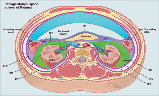
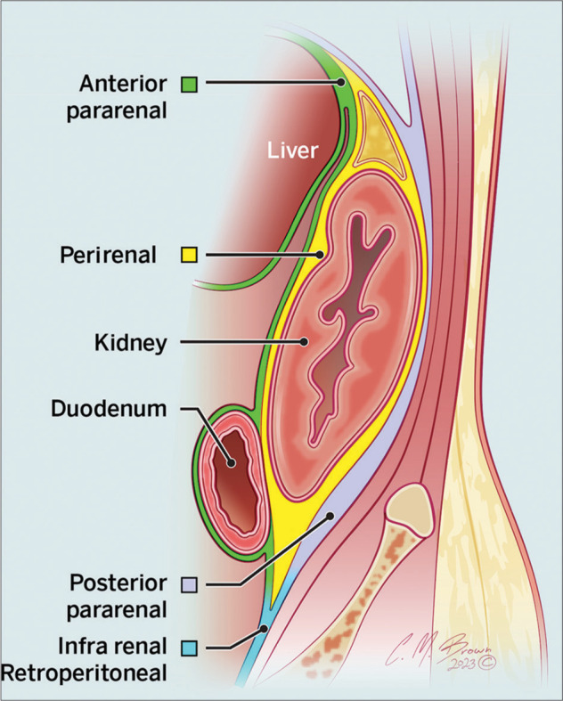
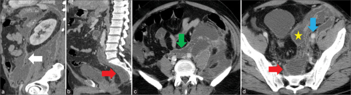
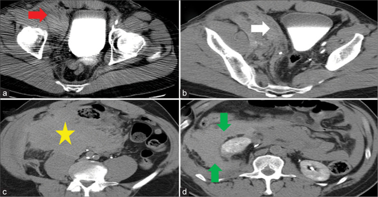
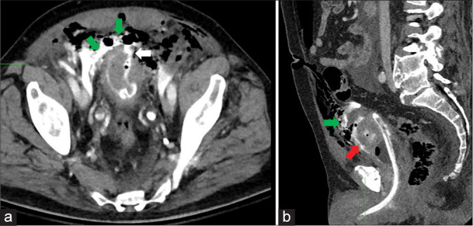
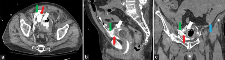
The retroperitoneum, a complex anatomical space within the abdominopelvic region, encompasses various vital abdominal organs. It is compartmentalized by fascial planes and contains potential spaces critical in multiple disease processes, including inflammatory effusions, hematomas, and neoplastic conditions. A comprehensive understanding of the retroperitoneum and its potential spaces is essential for radiologists in identifying and accurately describing the extent of abdominopelvic disease. This pictorial review aims to describe the anatomy of the retroperitoneum while discussing commonly encountered pathologies within this region. Through a collection of illustrative images, this review will provide radiologists with valuable insights into the retroperitoneum, facilitating their diagnostic proficiency to aid in appropriate patient clinical management.

Keywords: Anatomy; Extraperitoneal spaces; Pathologic fluid; Retroperitoneal spaces; Retroperitoneum.


Conflict of interest statement

There are no conflicts of interest.

Figures

Figure 1:
Figure 1:
Illustration of the retroperitoneum at the level of the kidneys demonstrating the retroperitoneal compartments (PP: Parietal peritoneum, APF: Anterior pararenal fascia, LCF: Lateroconal fascia, APS: Anterior pararenal space, PRS: Perirenal space, PPS: Posterior pararenal space, RRS: Retrorenal space, PPS, TF: Transversalis fascia).
Figure 2:
Figure 2:
Illustration of the retroperitoneal space at and below the level of kidneys.
Figure 3:
Figure 3:
A 40 year old woman with sepsis. Computed tomography (CT) axial image at the level of the kidneys demonstrates a perirenal abscess (star) contained by the anterior (white arrow) and posterior (red arrow) perirenal fascia.
Figure 4:
Figure 4:
A 49 year old man with blunt abdominal trauma. Computed tomography (CT) of the abdomen (a, c) axial, (b) sagittal reformatted images demonstrating injury to the bare area of the liver (white arrow) with superior perirenal hematoma (star) and adrenal hematoma (red arrow).
Figure 5:
Figure 5:
A 40 year old man with acute flank pain. Contiguous axial abdomen computed tomography (CT) images (a, b) demonstrating a left subcapsular urinoma/hemorrhage (star) secondary to obstructing stone (red arrow).
Figure 6:
Figure 6:
A 63 year old man with hemorrhagic pancreatitis. Computed tomography (CT) abdomen axial image demonstrating an abscess in the anterior pararenal space with extension into the lateral conal space (star).
Figure 7:
Figure 7:
A 65 year-old-man with complications from cardiac catheterization procedure. (a) Arterial angiogram and (b) axial computed tomography (CT) image of the abdomen demonstrating active extravasation from the right external iliac artery (red arrow). Hemorrhage is seen in the right anterior pararenal space (white arrow), which extends into the left anterior pararenal space (green arrow).
Figure 8:
Figure 8:
A 39 year old man with acute pancreatitis. Contiguous computed tomographic (CT) abdomen axial images demonstrating inflammatory fluid in the left anterior pararenal space (red arrow), which extends along the root of the mesentery and mesocolon (white arrows).
Figure 9:
Figure 9:
A 40 year old man with Pott Disease. Abdominal computed tomography (CT) (a) axial and (b) sagittal images demonstrating a large iliopsoas abscess with extension into the posterior pararenal space (star). Also noted is a destruction of an adjacent vertebral body (red arrow).
Figure 10:
Figure 10:
A 53 year old woman with pancreatitis. Abdominal computed tomography (CT) (a) axial and (b) sagittal images demonstrating inflammatory fluid in the posterior pararenal space (red arrow), which extends into the pelvic retroperitoneum (white arrow).
Figure 11:
Figure 11:
A 41 year old woman with perforated diverticulitis of the descending colon. Computed tomography (CT) abdomen axial image demonstrating air tracking in the retro mesenteric space (red arrow).
Figure 12:
Figure 12:
A 37 year old man with acute pancreatitis. Computed tomography (CT) abdomen axial image demonstrating inflammatory fluid in the lateral conal plane (red arrow) communicating with the retro mesenteric (white arrow) and retrorenal (star) spaces.
Figure 13:
Figure 13:
Illustration of the pelvic retroperitoneal spaces on sagittal and axial views.
Figure 14:
Figure 14:
A 40 year old man with acute pancreatitis. Contiguous (a and b) sagital reformat and (c and d) axial abdominopelvic images demonstrating an inflammatory fluid collection in the infrarenal space (white arrow) with extension into the pelvis prevesical (star) and presacral (red arrows) spaces. Both medial (green arrow) and iliac vascular (blue arrow) modes of spread are seen.
Figure 15:
Figure 15:
A 65 year old woman following right femoral catheterization. Contiguous abdominopelvic computed tomography (CT) axial images demonstrating a hematoma surrounding the femoral sheath 9 (red arrow). Hematoma is seen in the prevesical space (white arrow) with retrograde extension into the right infrarenal (star) and anterior and posterior pararenal spaces (green arrows).
Figure 16:
Figure 16:
A 69 year old man with blunt abdominal trauma. Pelvic computed tomography (CT) (a) axial and (b) sagittal images demonstrating injury to the anterior bladder wall with contrast seen in the perivesical (red arrow) and prevesical (green arrow) spaces via direct communication.
Figure 17:
Figure 17:
A 67 year old man with iatrogenic bladder injury during transurethral resection of the bladder tumor. Computed tomography (CT) cystogram (a) axial, (b) sagittal, and (c) coronal images demonstrating an injury to the anterosuperior bladder wall (white arrow). Contrast extravasation is seen with the perivesical (red arrow) and prevesical (green arrow) spaces. Contrast is seen with the large volume ascites (blue arrow) consistent with intraperitoneal urinoma.

Similar articles

References

    1. Tirkes T, Sandrasegaran K, Patel AA, Hollar MA, Tejada JG, Tann M, et al. Peritoneal and retroperitoneal anatomy and its relevance for cross-sectional imaging. Radiographics. 2012;32:437–51. doi: 10.1148/rg.322115032. - DOI - PubMed
    1. Coffin A, Boulay-Coletta I, Sebbag-Sfez D, Zins M. Radioanatomy of the retroperitoneal space. Diagn Interv Imaging. 2015;96:171–86. doi: 10.1016/j.diii.2014.06.015. - DOI - PubMed
    1. Quaia E, Gennari AG. Radiological imaging of the kidney. Berlin, Heidelberg: Springer Berlin Heidelberg; 2014. Normal radiological anatomy of the retroperitoneum; pp. 75–9. - DOI
    1. Lambert G, Samra NS. StatPearls. Treasure Island, FL: StatPearls Publishing; 2023. Anatomy, abdomen and pelvis, retroperitoneum. - PubMed
    1. Mindell HJ, Mastromatteo JF, Dickey KW, Sturtevant NV, Shuman WP, Oliver CL, et al. Anatomic communications between the three retroperitoneal spaces: Determination by CT-guided injections of contrast material in cadavers. AJR Am J Roentgenol. 1995;164:1173–8. doi: 10.2214/ajr.164.5.7717227. - DOI - PubMed

LinkOut - more resources