

Risk prediction of CISS classification in endovascular treatment of basilar artery stenosis
- PMID: 38205300
- PMCID: PMC10776930
- DOI: 10.1016/j.heliyon.2023.e23747
Risk prediction of CISS classification in endovascular treatment of basilar artery stenosis
Abstract
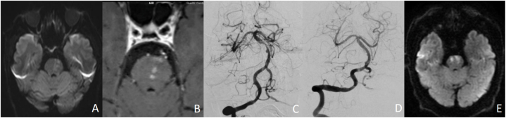
Objective: To investigate the incidence of ischemic stroke complications after endovascular treatment for basilar artery stenosis used preoperative high-resolution magnetic resonance vascular wall imaging (HRMR/VWI) and diffusion-weighted imaging (DWI).
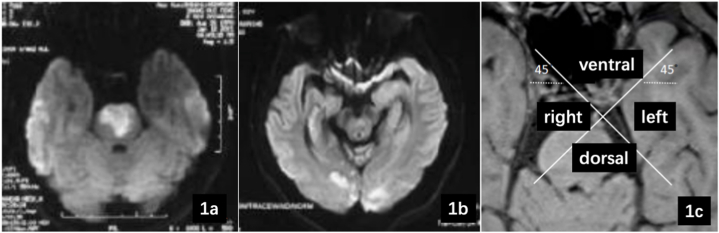
Methods: The clinical data of 47 patients with severe symptomatic basilar artery stenosis (stenosis rate ≥70 %) treated with endovascular therapy at the Neuro-interventional Center from December 2017 to December 2021 were retrospectively analyzed. High-resolution magnetic resonance angiography (HRMR VWI) and DWI were used to evaluate the location of atherosclerotic plaque at basilar artery stenosis and the distribution of cerebral infarction lesions in all patients before surgery.According to the CISS classification system for ischemic stroke, patients were divided into a perforation group and a hypoperfusion group, and the general situation, plaque distribution, and incidence of ischemic stroke complications 7 days after endovascular treatment in the two groups were analyzed.
Results: There was no significant difference in baseline data between the two groups. After 7 days of intravascular treatment, the incidence of ischemic stroke was higher in the perforation group (20.0 %) than in the hypoperfusion group (0.0 %), and the difference was statistically significant (P = 0.027). A significant association was found between the perforation group and the hypoperfusion group for the incidence of ischemic stroke at 7 days (P = 0.003, OR = 2.347; 95 % CI = 2.056-4.268). There were a higher proportion of ventral plaques (74.1 %) and a lower proportion of dorsal plaques (33.3 %) in the hypoperfusion group, which were 15.0 % and 90.0 % in the perforation group, respectively (χ2 = 16.045, P < 0.001; χ2 = 15.092, P < 0.001). There was no significant difference in the proportion of left and right plaques between the two groups.
Conclusions: The risk of ischemic stroke is greater in patients with perforator artery obstruction undergoing endovascular therapy, and lower in patients with hypoperfusion/embolus removal.
Keywords: Basilar artery stenosis; Endovascular therapy; High resolution magnetic resonance angiography; Plaque.
© 2023 The Authors. Published by Elsevier Ltd.
Conflict of interest statement
The authors declare that they have no known competing financial interests or personal relationships that could have appeared to influence the work reported in this paper.
Figures
References
-
- Wang Y.J., Li Z.X., Gu H.Q., Zhai Y., Zhou Q., Jiang Y., Zhao X.Q., Wang Y.L., Yang X., Wang C.J., et al. China stroke statistics: an update on the 2019 report from the national center for healthcare quality management in neurological diseases, China national clinical research center for neurological diseases, the Chinese stroke association, national center for chronic and non-communicable disease control and prevention, Chinese center for disease control and prevention and Institute for global neuroscience and stroke collaborations. Stroke Vasc Neurol. 2022;7(5):415–450. doi: 10.1136/svn-2021-001374. - DOI - PMC - PubMed
-
- Abuzinadah A.R., Alanazy M.H., Almekhlafi M.A., Duan Y., Zhu H., Mazighi M., Lutsep H.L., Donnon T., Hill M.D. Stroke recurrence rates among patients with symptomatic intracranial vertebrobasilar stenoses: systematic review and meta-analysis. J. Neurointerventional Surg. 2016;8(2):112–116. doi: 10.1136/neurintsurg-2014-011458. - DOI - PMC - PubMed
-
- Li K., Sun D., Tong X., Wang A., Zhang Y., Ma G., Huo X., Ma N., Gao F., Mo D., et al. Incidence, predictors, and impact on outcome of underlying intracranial atherosclerotic disease in acute vertebrobasilar artery occlusion undergoing endovascular therapy: data from ANGEL-ACT registry. Int. J. Stroke. 2023 doi: 10.1177/17474930221150111. - DOI - PubMed
LinkOut - more resources
Full Text Sources
