. 2023 Aug 16;8(12):1539-1554.
doi: 10.1016/j.jacbts.2023.05.010. eCollection 2023 Dec.

Extracellular Matrix Protein-1 as a Mediator of Inflammation-Induced Fibrosis After Myocardial Infarction

Affiliations

Extracellular Matrix Protein-1 as a Mediator of Inflammation-Induced Fibrosis After Myocardial Infarction

Sean A Hardy et al. JACC Basic Transl Sci. .

Abstract

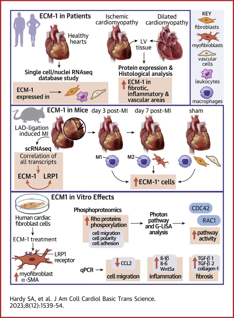
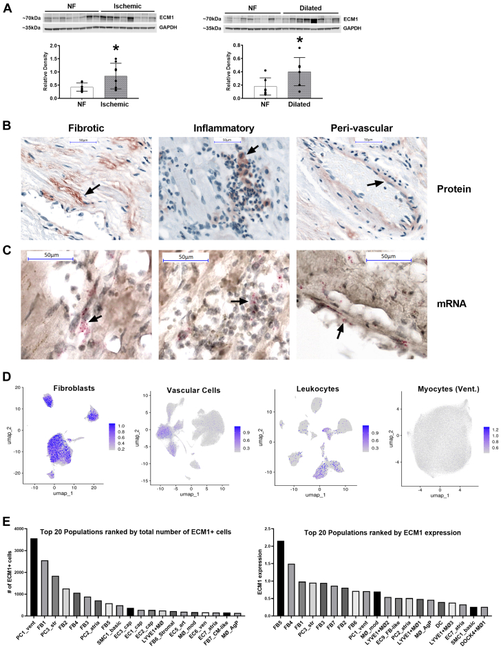
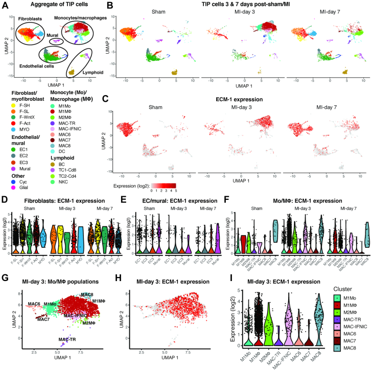
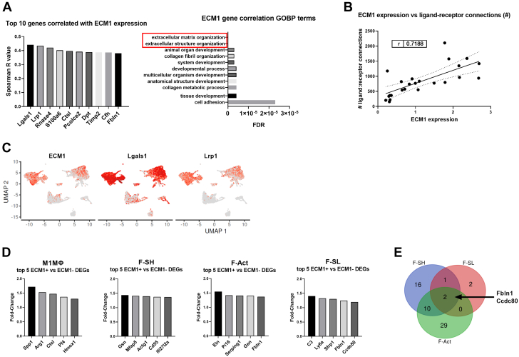
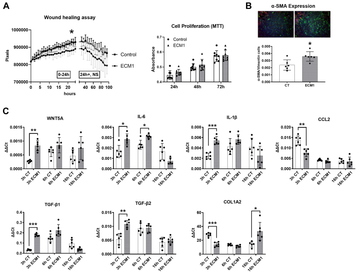
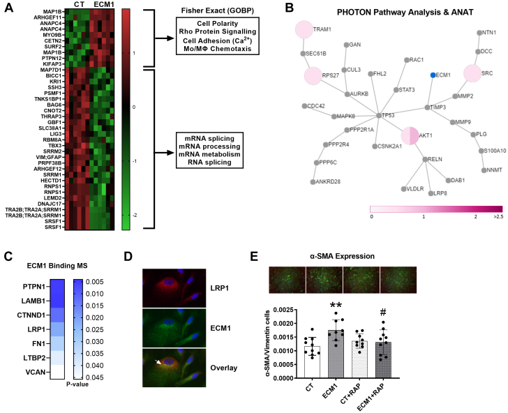
Irreversible fibrosis is a hallmark of myocardial infarction (MI) and heart failure. Extracellular matrix protein-1 (ECM-1) is up-regulated in these hearts, localized to fibrotic, inflammatory, and perivascular areas. ECM-1 originates predominantly from fibroblasts, macrophages, and pericytes/vascular cells in uninjured human and mouse hearts, and from M1 and M2 macrophages and myofibroblasts after MI. ECM-1 stimulates fibroblast-to-myofibroblast transition, up-regulates key fibrotic and inflammatory pathways, and inhibits cardiac fibroblast migration. ECM-1 binds HuCFb cell surface receptor LRP1, and LRP1 inhibition blocks ECM-1 from stimulating fibroblast-to-myofibroblast transition, confirming a novel ECM-1-LRP1 fibrotic signaling axis. ECM-1 may represent a novel mechanism facilitating inflammation-fibrosis crosstalk.

Keywords: extracellular matrix; fibroblasts; fibrosis; heart; inflammation; myocardial infarction.


Conflict of interest statement

This work was supported by the Austrian Society of Cardiology, ERA-CVD, and Austrian Science Fund AIR-MI consortium (I 4168-B to Dr Rainer); John Hunter Charitable Trust (G1800510 to Dr Boyle); Hunter Medical Research Institute and Emlyn and Jennie Thomas Postgraduate Medical Research Scholarship (G2100164 and G1800696 to Dr Boyle); Australian Commonwealth funded Research Training Program stipend (to Drs Hardy and Mabotuwana); Medical University of Graz Doctoral School of Translational Molecular and Cellular Biosciences [to Drs Hardy and Mabotuwana] and Doctoral School of Molecular Medicine [to Dr Rech]; Austrian Society of Cardiology (to Dr Hardy); National Health and Medical Research Council (NHMRC) (2000615 and 1074386 to Dr Harvey, 1079187 and 1175134 to Dr Hansbro, and 1156898 and 20000483 to Dr Starkey); NHMRC Senior Principal Research Fellowship (1118576 to Dr Harvey); Stem Cells Australia (SR110001002 to Dr Harvey); the Victor Chang Cardiac Research Institute, Australia [to Dr Harvey]; the University of Technology Sydney, Australia [to Dr Hansbro]; Monash University, Australia (to Dr Starkey); Australian Research Council (DE170100226 to Dr Starkey); Foundation Leducq (15CVD03 and 13CVD01 to Dr Harvey); Austrian Science fund (KLI645, W1226, and F73 to Dr Birner-Gruenberger); Interdisciplinary Centre for Clinical Research, University Hospital Würzburg (E-353 to Dr Cochain, E-354 to Dr Campos Ramos); the German Research Foundation [471705758 and 458539578 to Dr Cochain, DFG SFB1525 project no. 453989101 to Drs Cochain and Campos Ramos, and 411619907 to Dr Campos Ramos]; and the European Research Area Network-Cardiovascular Diseases/German Federal Ministry of Education and Research (01KL1902 to Dr Campos Ramos). All other authors have reported that they have no relationships relevant to the contents of this paper to disclose.PerspectivesCOMPETENCY IN MEDICAL KNOWLEDGE: Fibrosis is a key factor in heart disease that impedes cardiac function and prognosis. Yet, no therapies specifically target profibrotic signaling in the heart, and once established, fibrotic remodeling is largely irreversible. Inflammation-fibrosis crosstalk, whereby ECM remodeling and fibrotic tissue deposition is tightly connected to inflammation and vice versa, is now thought to be critical in orchestrating wound healing and cardiac scarring. It is suggested that this is why unidirectional therapies targeting fibrosis or inflammation alone have failed to substantially reduce the mortality associated with cardiac diseases. Here, we represent ECM-1 as a potential novel mediator of inflammation-induced fibrotic signaling in the heart and confirm its regulation in human heart failure. TRANSLATIONAL OUTLOOK: ECM-1 may serve as an attractive future treatment target to prevent excessive and detrimental fibrosis. Because fibrosis and scarring is a universal response to injury in many organs, this has potential implications beyond heart disease.

Figures

None
Graphical abstract
Figure 1
Figure 1
ECM-1 Is Upregulated in Human Failing Hearts and Is Localized to Fibrotic Tissue, Leukocytes, and Endothelia (A) Western blotting shows that extracellular matrix protein (ECM)-1 protein is up-regulated in ischemic and dilated human heart failure patients, presented as mean ± SD, analyzed by means of Student’s t-test (n = 8/group). ∗P < 0.05. (B) Immunohistochemistry of ECM-1 protein expression in the human heart (NF: n = 4; heart failure: n = 8) shows that ECM-1 expression is predominantly interstitial, localized to fibrotic, inflammatory, and perivascular areas (×40 magnification; scale bars = 50 μm). (C) mRNA in situ hybridization (n = 3) confirms transcription of ECM-1 in these areas (×40 magnification zoomed in; scale bars = 50 μm). (D) ECM-1-specific analysis of single-cell/single-nuclear RNA sequencing dataset from Litviňuková et al identifies ECM-1 expression by fibroblasts, leukocytes, and vascular cells in the healthy human heart (gray = all cells; blue overlay = ECM-1+ cells). (E) The top 20 ECM-1-expressing cell subpopulations ranked by total number of ECM-1+ cells and by ECM-1 expression level (expressed as a Log2 average). NF = nonfailing.
Figure 2
Figure 2
Expression of ECM-1 in Cardiac Interstitial Cells Before and After MI (A) UMAP plot showing cell populations in single-cell RNA sequencing of cardiac interstitial cells from sham-operated and MI hearts combined, and (B) faceted according to condition. (C) ECM-1 expression on UMAP coordinates according to condition. (D, E, F) Violin plots of ECM-1 expression according to condition for cell subpopulations in (D) fibroblasts, (E) endothelial (EC) and mural cells, and (F) monocyte (Mo) and macrophage (MΦ) populations. (G) UMAP plot of Mo/MΦ cell populations at day 3 after MI. (H, I) ECM-1 expression in day-3 post-MI Mo/MΦ cells on (H) UMAP coordinates and (I) in violin plots.Abbreviations as in Figure 1.
Figure 3
Figure 3
ECM-1 Expression Strongly Correlated With the Number of Outbound Ligand-Receptor Interactions Between Cells, Extracellular Matrix Organization, and Cell Adhesion Genes (A) Per-cell Spearman correlation analysis of ECM-1 expression against all genes in the mouse single-cell RNA sequencing data set; all P <0.001. The top 10 positively correlated genes (x-axis) are shown as column graphs of Spearman’s r-value (y-axis). The top ECM-1-correlated genes (with Spearman’s r > 0.30) were subject to gene ontology biological process (GOBP) enrichment analysis via the GOnet/DICE online tool. The top 10 GOBPs are presented as a bar chart ranked by false discovery rate (FDR) P value. (B) The average ECM-1 expression level (Log2) of each cell subpopulation is positively correlated with the number of significant outbound ligand-receptor connections (r = 0.719; P < 0.001), analyzed by Pearson’s correlation and simple linear regression. (C) ECM-1, Lgals1, and Lrp1 expression overlayed on the UMAP plot of cardiac interstitial cells from sham and MI (as shown in Figure 2A), to compare the cellular expression profile of the top 2 ECM-1 correlated genes. (D) Differentially expressed genes (DEGs) between ECM-1+ and ECM-1− cells (per cell subpopulation) within the major ECM-1-expressing cell subpopulations were assessed via MAST. Only M1MΦ, F-Act, F-SH, and F-SL returned lists of significant DEGs, and the top 5 up-regulated genes in ECM-1+ cells (x-axis) are shown in column graphs as fold change in gene expression (y-axis). (E) All up-regulated DEGs for M1MΦ, F-Act, F-SH, and F-SL were subject to Venn diagram analysis. No genes were commonly up-regulated in all 4 cell types; Fbln1 and Ccdc2 were commonly up-regulated in ECM-1+ F-Act, F-SH, and F-SL cells. Abbreviations as in Figures 1 and 2.
Figure 4
Figure 4
ECM-1 Inhibits Human Cardiac Fibroblast Cell Migration in Culture, and Stimulates Fibroblast-to-Myofibroblast Transition and Inflammatory and Fibrotic Signaling Pathways in HuCFbs (A) ECM-1 treatment (20 ng/mL) significantly reduced the migration rate of HuCFb cells over 24 hours relative to control subjects (n = 4/group), as assessed via both repeated-measures 2-way analysis of variance (ANOVA) with post hoc Šídák’s multiple comparisons test (P = 0.044) and inclination of the linear equation of growth rate via Student’s t-test (P = 0.027). ECM-1 had no effect on cell proliferation assessed via MTT assay after 24, 48, or 72 hours of treatment, as assessed via repeated-measures 2-way ANOVA with post hoc Šídák’s multiple comparisons test. (B) Immunofluorescence shows ECM-1 stimulates fibroblast-to-myofibroblast transition in HuCFbs after 48 hours of treatment, as analyzed by Student’s t-test (P = 0.014; n = 6; ×2.5 magnification; green = α-smooth muscle actin [SMA]; red = vimentin; blue = DAPI/nuclei). (C) HuCFbs were treated with ECM-1 (20 ng/mL) or medium alone for 3, 6, or 16 hours and assessed via quantitative polymerase chain reaction for mRNA expression of Wnt5a, interleukin (IL)-1β2, IL-6, CCL2, transforming growth factor (TGF)-β1, TGF-β2, and collagen type I alpha 2 chain (Col1a2). Each gene was assessed via repeated-measures 2-way ANOVA (IL-6 and Col1a2 were assessed via repeated-measures mixed-effects analysis to account for missing values) with post hoc Šídák’s multiple comparisons test; expression of all genes are presented as the delta-delta threshold cycle (ΔΔCt) relative to Tpt1 housekeeping gene expression, n = 6/group. ∗P < 0.05; ∗∗P < 0.01; ∗∗∗P < 0.001. CT = control; other abbreviations as in Figures 1 and 2.
Figure 5
Figure 5
Proteomics Reveals ECM-1-HuCFb Cell Membrane Binding Partners and Downstream Signaling Mechanisms: ECM-1 Binds LRP1 Cell Surface Receptor, Which Is Required for ECM-1-Dependent Stimulation Of Profibrotic Fibroblast-to-Myofibroblast Transition (A) HuCFb cells were treated with ECM-1 (20 ng/mL) for 10 minutes and subject to phosphoproteomics (Ser/Thr) mass spectrometry (titanium dioxide workflow; n = 6/group). Thirty-eight proteins with significantly different phosphorylation levels were identified between CT (medium alone) and ECM-1-treated cells, as assessed via Student’s t-test with permutation-based FDR; data are presented as the Log2 difference (CT − ECM-1) in phosphorylation level; green = down-regulated; red = up-regulated. Note that proteins measured multiple times represent different phosphosites within those proteins. Fisher’s exact enrichment testing was conducted on significantly up-regulated and down-regulated phosphorylation sites in ECM-1-treated samples (separately), and 4 of the most over-represented GOBPs are shown. (B) Our list of differential phosphorylation sites in ECM-1-treated HuCFbs was then subjected to PHOTON pathway analysis (2-sided) with reconstruction ANAT (ECM-1 as the response source) to visualize the ECM-1-dependent signaling network. The ANAT network for ECM-1 signaling in the “greater” PHOTON analysis direction (signaling score ≥6) is shown with ECM-1 highlighted in blue. All proteins shown in the network represent involvement in active mediation of ECM-1-dependent signal transduction. Pink pie charts represent fold changes of measured phosphorylation sites (see scale bar), with multiple phosphorylation sites represented by a pie chart divided by the number of phosphorylation sites measured. (C) ECM-1 binding partners were pulled down from a purified HuCFb membrane protein lysate, with the use of recombinant human ECM-1 protein as bait; 58 potential ECM-1-binding proteins were identified and 7 proteins of particular interest are presented: PTPN1, LAMB1, CTNND1, LRP1, FN1, LTBP2, and VCAN. (D) Immunofluorescence of HuCFb cells in culture showed that ECM-1 colocalizes with LRP1 (×40 magnification zoomed in); arrowhead shows an example area of interest (red = low-density lipoprotein receptor–related protein 1 [LRP1]; green = ECM-1; blue = DAPI/nuclei). (E) Immunofluorescence shows ECM-1 stimulates fibroblast-to-myofibroblast transition in HuCFbs at 48 hours of treatment (P = 0.003), and RAP (LRPAP1)–dependent LRP1 inhibition blocks this effect (ECM-1 vs ECM-1+RAP: P = 0.038; CT vs ECM-1+RAP; nonsignificant), presented as mean ± SD, analyzed by 1-way ANOVA with post hoc Šídák’s multiple comparisons test (n ≥ 9; ×2.5 magnification; green = α-SMA; red = vimentin; blue = DAPI/nuclei); #P < 0.05 comparing ECM-1+RAP and ECM-1 treatment groups. ∗P < 0.05; ∗∗P < 0.01. Abbreviations as in Figure 1, Figure 2, Figure 3, Figure 4.

References

    1. Hardy S.A., Mabotuwana N.S., Murtha L.A., et al. Novel role of extracellular matrix protein 1 (ECM1) in cardiac aging and myocardial infarction. PLoS One. 2019;14 - PMC - PubMed
    1. Sansbury B.E., DeMartino A.M., Xie Z., et al. Metabolomic analysis of pressure-overloaded and infarcted mouse hearts. Circ Heart Fail. 2014;7:634–642. - PMC - PubMed
    1. Forte E., Furtado M.B., Rosenthal N. The interstitium in cardiac repair: role of the immune–stromal cell interplay. Nat Rev Cardiol. 2018;15:601–616. - PubMed
    1. Kretzschmar K., Post Y., Bannier-Hélaouët M., et al. Profiling proliferative cells and their progeny in damaged murine hearts. Proc Natl Acad Sci U S A. 2018;115 - PMC - PubMed
    1. Sadek H., Olson E.N. Toward the goal of human heart regeneration. Cell Stem Cell. 2020;26:7–16. - PMC - PubMed