Review
. 2023 Jun 21;8(12):1613-1628.
doi: 10.1016/j.jacbts.2023.03.024. eCollection 2023 Dec.

The Cardiac Circadian Clock: Implications for Cardiovascular Disease and its Treatment

Affiliations
Review

The Cardiac Circadian Clock: Implications for Cardiovascular Disease and its Treatment

Martin E Young. JACC Basic Transl Sci. .

Abstract

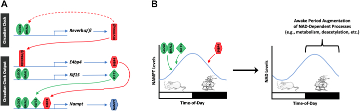
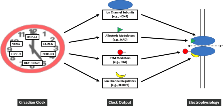
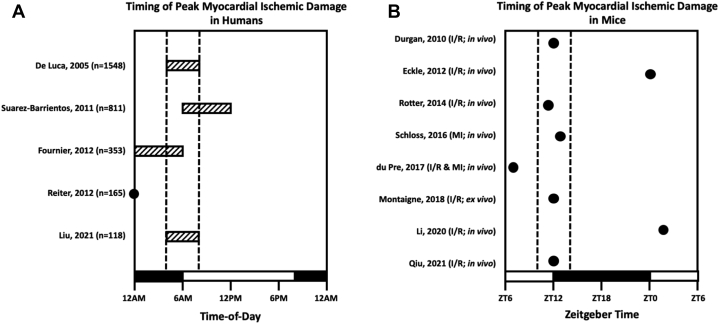
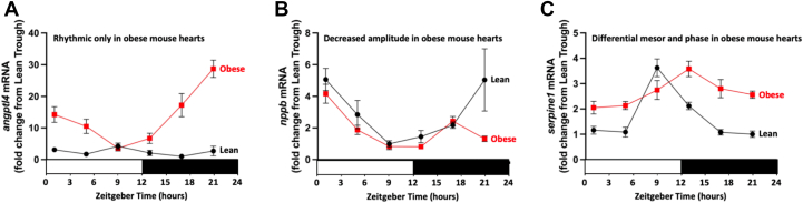
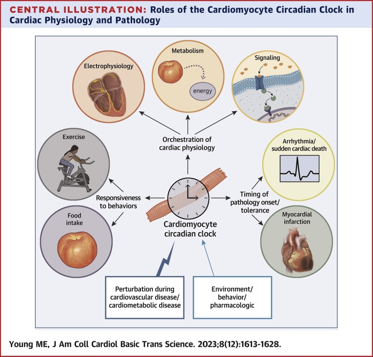
Virtually all aspects of physiology fluctuate with respect to the time of day. This is beautifully exemplified by cardiovascular physiology, for which blood pressure and electrophysiology exhibit robust diurnal oscillations. At molecular/biochemical levels (eg, transcription, translation, signaling, metabolism), cardiovascular-relevant tissues (such as the heart) are profoundly different during the day vs the night. Unfortunately, this in turn contributes toward 24-hour rhythms in both risk of adverse event onset (eg, arrhythmias, myocardial infarction) and pathogenesis severity (eg, extent of ischemic damage). Accumulating evidence indicates that cell-autonomous timekeeping mechanisms, termed circadian clocks, temporally govern biological processes known to play critical roles in cardiovascular function/dysfunction. In this paper, a comprehensive review of our current understanding of the cardiomyocyte circadian clock during both health and disease is detailed. Unprecedented basic, translational, and epidemiologic studies support a need to implement chronobiological considerations in strategies designed for both prevention and treatment of cardiovascular disease.

Keywords: chronobiology; electrophysiology; heart failure; ischemia; metabolism.


Conflict of interest statement

This work was supported by the National Heart, Lung, and Blood Institute (HL149159 and HL007081). Dr Young has reported that he has no relationships relevant to the contents of this paper to disclose.

Figures

None
Graphical abstract
Figure 1
Figure 1
Clock Control of Cardiac NAD Levels (A) Integral circadian clock components BMAL1, CLOCK, and REV-ERBα/β form a negative feedback loop of the timekeeping mechanism. The BMAL1/CLOCK heterodimer induces transcription of the clock-controlled gene Krüppel-like factor 15 (KLF15), while REV-ERBα/β represses expression of the clock-controlled gene E4 promoter binding protein 4 (E4BP4). The clock-controlled gene nicotinamide phosphoribosyltransferase (NAMPT) (a key enzyme in the nicotinamide adenine dinucleotide [NAD] salvage pathway) is induced by both the BMAL1/CLOCK heterodimer and KLF15 and is repressed by E4BP4. (B) During the sleep phase, the BMAL1/CLOCK heterodimer and KLF15 induce NAMPT (peak activation at the sleep-to-wake transition), whereas E4BP4 represses NAMPT during the awake phase (peak repression at the wake-to-sleep transition). Twenty-four-hour oscillations in NAMPT lead to augmentation of cardiac NAD levels during the awake period, thereby facilitating NAD-dependent processes at this time. Green represents activation; red represents inhibition.
Figure 2
Figure 2
Direct and Indirect Regulation of Cardiac Electrophysiology by the Cardiomyocyte Circadian Clock Evidence-based hypothetical model depicting clock control of electrophysiology through alterations in ion channel subunits, allosteric modulators, mediators of post-translational modifications (PTMs), and regulatory proteins. CRY = cryptochrome; NAD = nicotinamide adenine dinucleotide; NAMPT = nicotinamide phosphoribosyltransferase; PER, period; PKA = protein kinase A.
Figure 3
Figure 3
Time of Day–Dependent Fluctuations in Myocardial Ischemic Tolerance (A) Correlation of greatest myocardial damage (based on circulating damage markers) with estimated timing of ischemia onset in humans. Dashed lines represent the 4-hour window during which peak myocardial damage clusters in the majority of reported studies (4 am to 8 am [ie, the sleep-to-wake transition]). (B) Correlation of greatest myocardial damage (based on infarct size) with known time of ischemic onset in mice. Zeitgeber time 0 (ZT0) is the time at which lights turn on in an animal facility, while ZT12 is the time at which lights turn off. Given that rodents are nocturnal, ZT12 represents the sleep-to-wake transition. Dashed lines represent the 4-hour window during which peak myocardial damage clusters in the majority of reported studies (ZT10-14 [ie, the sleep-to-wake transition]). I/R = ischemia/reperfusion; MI = myocardial infarction.
Figure 4
Figure 4
Differential 24-Hour Oscillations in Key Cardiokines During Obesity Time of day–dependent oscillations in cardiac (A) Angptl4, (B) Nppb, and (C) Serpine1 messenger RNA (mRNA) are altered to differing extents in obese mice compared with lean mice. Obesity was induced in mice through 20 weeks of high-fat feeding. Figures were drawn using publicly available data.
Central Illustration
Roles of the Cardiomyocyte Circadian Clock in Cardiac Physiology and Pathology The cardiomyocyte circadian clock temporally orchestrates processes that are critical for normal cardiac function (eg, electrophysiology, metabolism, and signaling). In doing so, the cardiomyocyte circadian clock influences responsiveness of the heart to both physiologic stimuli (eg, exercise and nutrients) as well as pathologic stresses (eg, myocardial infarction). In order to maintain its physiologic advantage, the cardiomyocyte circadian clock is exquisitely sensitive to environmental cues (which are perturbed during common cardiometabolic/cardiovascular disease states).

Similar articles

Cited by

References

    1. Degaute J.P., van de Borne P., Linkowski P., Van Cauter E. Quantitative analysis of the 24-hour blood pressure and heart rate patterns in young men. Hypertension. 1991;18(2):199–210. - PubMed
    1. Black N., D’Souza A., Wang Y., et al. Circadian rhythm of cardiac electrophysiology, arrhythmogenesis, and the underlying mechanisms. Heart Rhythm. 2019;16(2):298–307. - PMC - PubMed
    1. Voutilainen S., Kupari M., Hippelainen M., Karppinen K., Ventila M. Circadian variation of left ventricular diastolic function in healthy people. Heart. 1996;75(1):35–39. - PMC - PubMed
    1. Muller J., Tofler G., Stone P. Circadian variation and triggers of onset of acute cardiovascular disease. Circulation. 1989;79:733–743. - PubMed
    1. Prinz P.N., Halter J., Benedetti C., Raskind M. Circadian variation of plasma catecholamines in young and old men: relation to rapid eye movement and slow wave sleep. J Clin Endocrinol Metab. 1979;49(2):300–304. - PubMed

LinkOut - more resources