. 2023 Dec 27:14:1213726.
doi: 10.3389/fphar.2023.1213726. eCollection 2023.

A glycoengineered therapeutic anti-HBV antibody that allows increased HBsAg immunoclearance improves HBV suppression in vivo

Affiliations

A glycoengineered therapeutic anti-HBV antibody that allows increased HBsAg immunoclearance improves HBV suppression in vivo

Min You et al. Front Pharmacol. .

Abstract

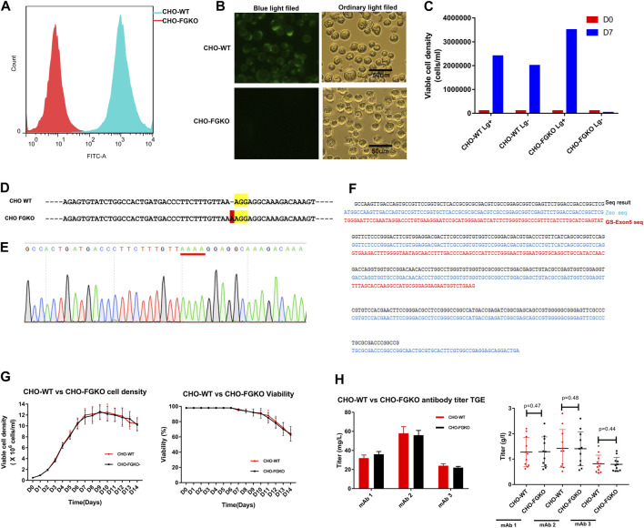
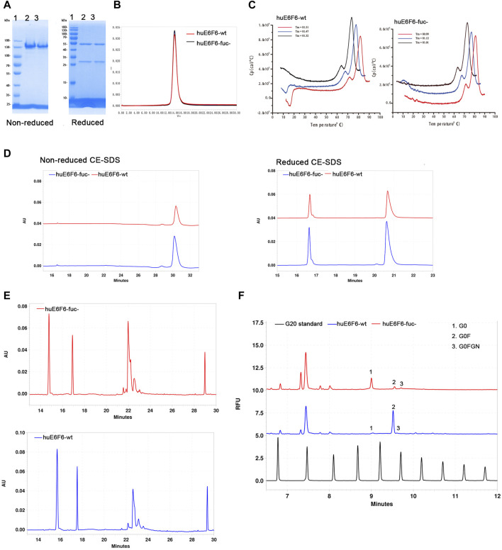
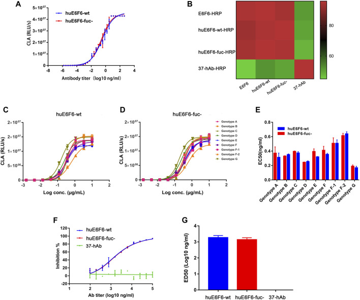
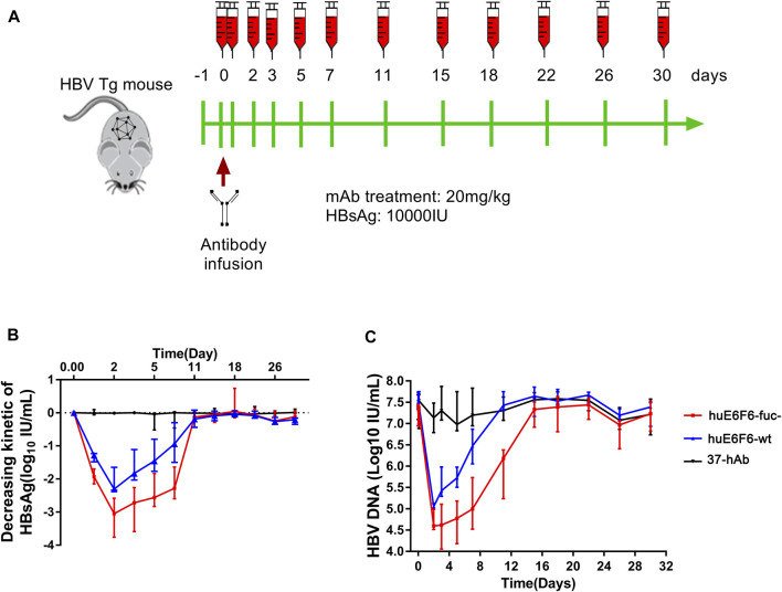
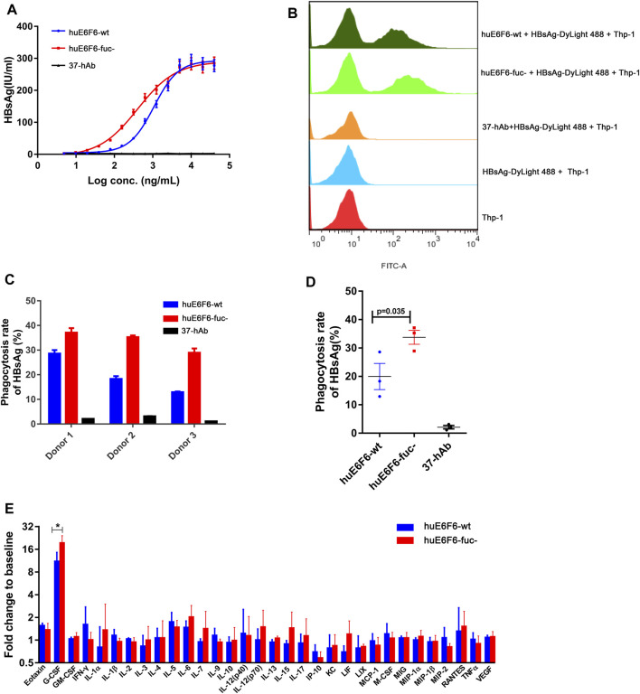
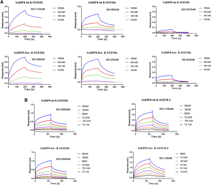
Introduction: The effective and persistent suppression of hepatitis B surface antigen (HBsAg) in patients with chronic HBV infection (CHB) is considered to be a promising approach to achieve a functional cure of hepatitis B. In our previous study, we found that the antibody E6F6 can clear HBsAg through FcγR-mediated phagocytosis, and its humanized form (huE6F6 antibody) is expected to be a new tool for the treatment of CHB. Previous studies have shown that the glycosylation of Fc segments affects the binding of antibodies to FcγR and thus affects the biological activity of antibodies in vivo. Methods: To further improve the therapeutic potential of huE6F6, in this study, we defucosylated huE6F6 (huE6F6-fuc-), preliminarily explored the developability of this molecule, and studied the therapeutic potential of this molecule and its underlying mechanism in vitro and in vivo models. Results: huE6F6-fuc- has desirable physicochemical properties. Compared with huE6F6-wt, huE6F6-fuc- administration resulted in a stronger viral clearance in vivo. Meanwhile, huE6F6-fuc- keep a similar neutralization activity and binding activity to huE6F6-wt in vitro. Immunological analyses suggested that huE6F6-fuc- exhibited enhanced binding to hCD32b and hCD16b, which mainly contributed to its enhanced therapeutic activity in vivo. Conclusions: In summary, the huE6F6-fuc- molecule that was developed in this study, which has desirable developability, can clear HBsAg more efficiently in vivo, providing a promising treatment for CHB patients. Our study provides new guidance for antibody engineering in other disease fields.

Keywords: HBsAg; chronic hepatitis B infection; defucosylation; neutralization; phagocytosis; therapeutic antibody.


Conflict of interest statement

The authors declare that the research was conducted in the absence of any commercial or financial relationships that could be construed as a potential conflict of interest. The reviewer (CS) declared a past co-authorship with the authors (WL, YC, XL, CF, XW, BZ, MF, TZ, YQ) to the handling editor

Figures

FIGURE 1
FIGURE 1
Development of FUT8−/−GS−/− knockout CHO cells. (A) CHO-WT and CHO-FGKO cells stained with lectin- FITC were analyzed by FACS. (B) CHO-WT and CHO-FGKO cells were stained with Lectin-FITC. (C) CHO-WT and CHO-FGKO cells were cultured with or without Lg. (D) Exon 7 of FUT8 in CHO-WT and CHO-FGKO were aligned. The nucleotides in yellow are PAM sites, and the nucleotides in red are insertions. (E) Sanger sequencing of exon 7 of CHO-FGKO cells is shown. (F) GS Exon 5 of CHO-FGKO cells was sequenced and assayed. (G) CHO-WT and CHO-FGKO cells were cultured with a fed-batch approach. The cell density is shown on the left, and the cell viability is shown on the right. (H) CHO-WT and CHO-FGKO cells were used for the expression of antibodies. The left figure shows the transient expression yield, and the stable cell pool expression yield of the three antibodies (mAb1, mAb2 and mAb3) is shown on the right.
FIGURE 2
FIGURE 2
Physicochemical properties of huE6F6-wt and huE6F6-fuc-. (A) SDS‒PAGE analysis of the antibodies under nonreduced or reduced conditions. Lane 1, protein markers; lane 2, huE6F6-wt; lane 3, huE6F6-fuc-. (B) SEC assay of huE6F6-wt and huE6F6-fuc-. (C) DSC assay of huE6F6-wt and huE6F6-fuc- in PBS. (D) CE-SDS analysis of huE6F6-wt and huE6F6-fuc- under nonreduced or reduced conditions. (E) The ciEF assay of huE6F6-wt and huE6F6-fuc-. (F) The N-Glycan assay of huE6F6-wt and huE6F6-fuc- with PA800.
FIGURE 3
FIGURE 3
Binding and neutralizing activity of antibodies in vitro. (A) The binding activity of huE6F6-wt and huE6F6-fuc- mAbs with HBsAg. (B) Competition between antibodies. The competition level is shown in color. E6F6 is the parent molecule of huE6F6-wt and huE6F6-fuc-. 37-hAb, which is a humanized antibody against H5N1, was used as an isotype control. (C) Binding of huE6F6-wt to peptide epitopes from all HBV genotypes. (D) Binding of huE6F6-fuc- to peptide epitopes from all HBV genotypes. (E) The EC50 of huE6F6-wt or huE6F6-fuc- binding the epitope of the different HBV genotypes. (F) The neutralization activity of huE6F6-wt and huE6F6-fuc- mAbs against HBV. (G) The ED50s were calculated according to Figure 3F.
FIGURE 4
FIGURE 4
In vivo suppression of HBV by huE6F6-fuc-. (A) Schematic diagram showing the bleeding and antibody infusion schedules in the mouse study. Five animals were included in each group, except that three were included in the 37-hAb group. (B) Decreasing kinetics of HBsAg in HBV-Tg mice after a single mAb treatment. (C) HBV DNA concentrations in the mice.
FIGURE 5
FIGURE 5
Defucosylation enhances antigen phagocytosis. (A) The level of antibody-mediated internalization of HBsAg by differentiated THP-1 cells. (B) Flow cytometric analyses of the mAb-HBsAg ICs in differentiated THP-1 cells. (C) The huE6F-mediated phagocytosis of HBsAg by PBMCs. (D) Scatter plot analysis of phagocytosis efficiency by PBMCs. (E) Serum cytokine profile of HBV-Tg mice (n = 3) that received mAbs at 6 h after infusion.
FIGURE 6
FIGURE 6
Representative BLI analysis of huE6F6-wt or huE6F6-fuc- binding to Fcγ receptors. (A) Kinetics of huE6F6-wt or huE6F6-fuc- binding to hCD32b, hCD16a, and hCD16b. (B) Kinetics of huE6F6-wt or huE6F6-fuc- binding to mCD32b and mCD16-2.

References

    1. Bakalar M. H., Joffe A. M., Schmid E. M., Son S., Podolski M., Fletcher D. A. (2018). Size-dependent segregation controls macrophage phagocytosis of antibody-opsonized targets. Cell 174, 131–142. 10.1016/j.cell.2018.05.059 - DOI - PMC - PubMed
    1. Barouch D. H., Whitney J. B., Moldt B., Klein F., Oliveira T. Y., Liu J., et al. (2013). Therapeutic efficacy of potent neutralizing HIV-1-specific monoclonal antibodies in SHIV-infected rhesus monkeys. Nature 503, 224–228. 10.1038/nature12744 - DOI - PMC - PubMed
    1. Baum A., Ajithdoss D., Copin R., Zhou A., Lanza K., Negron N., et al. (2020). REGN-COV2 antibodies prevent and treat SARS-CoV-2 infection in rhesus macaques and hamsters. Science 370, 1110–1115. 10.1126/science.abe2402 - DOI - PMC - PubMed
    1. Bournazos S. (2019). IgG Fc receptors: evolutionary considerations. Curr. Top. Microbiol. Immunol. 423, 1–11. 10.1007/82_2019_149 - DOI - PubMed
    1. Bruhns P., Jonsson F. (2015). Mouse and human FcR effector functions. Immunol. Rev. 268, 25–51. 10.1111/imr.12350 - DOI - PubMed

LinkOut - more resources