

Central role of podocytes in mediating cellular cross talk in glomerular health and disease
- PMID: 38205544
- PMCID: PMC11207540
- DOI: 10.1152/ajprenal.00328.2023
Central role of podocytes in mediating cellular cross talk in glomerular health and disease
Abstract
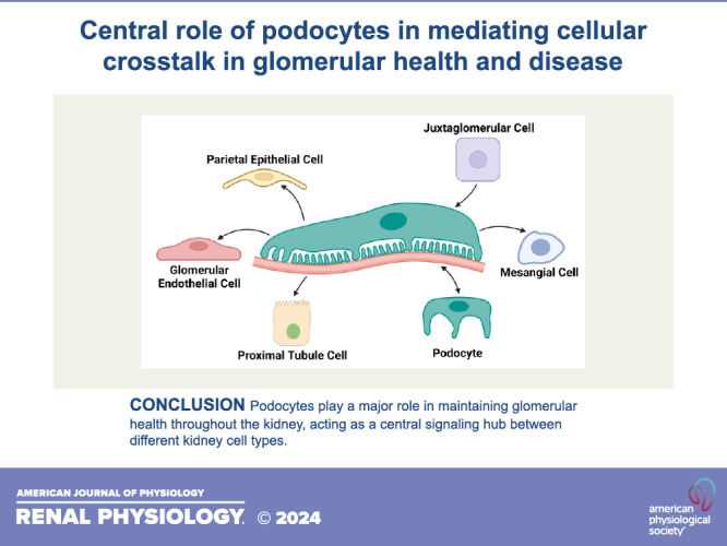
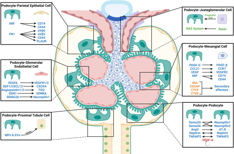
Podocytes are highly specialized epithelial cells that surround the capillaries of the glomeruli in the kidney. Together with the glomerular endothelial cells, these postmitotic cells are responsible for regulating filtrate from the circulating blood with their organized network of interdigitating foot processes that wrap around the glomerular basement membrane. Although podocyte injury and subsequent loss is the hallmark of many glomerular diseases, recent evidence suggests that the cell-cell communication between podocytes and other glomerular and nonglomerular cells is critical for the development and progression of kidney disease. In this review, we highlight these key cellular pathways of communication and how they might be a potential target for therapy in glomerular disease. We also postulate that podocytes might serve as a central hub for communication in the kidney under basal conditions and in response to cellular stress, which may have implications for the development and progression of glomerular diseases.
Keywords: cellular cross talk; glomerular diseases; kidney disease; podocytes.
Conflict of interest statement
Sandeep K. Mallipattu is an editor of the
Figures
Similar articles
-
Glomerular Endothelium and its Impact on Glomerular Filtration Barrier in Diabetes: Are the Gaps Still Illusive?Curr Med Chem. 2018;25(13):1525-1529. doi: 10.2174/0929867324666170705124647. Curr Med Chem. 2018. PMID: 28685678 Review.
-
Podocyte injury and its consequences.Kidney Int. 2016 Jun;89(6):1221-30. doi: 10.1016/j.kint.2016.01.012. Epub 2016 Mar 19. Kidney Int. 2016. PMID: 27165817 Review.
-
Glomerular endothelial derived vesicles mediate podocyte dysfunction: A potential role for miRNA.PLoS One. 2020 Mar 26;15(3):e0224852. doi: 10.1371/journal.pone.0224852. eCollection 2020. PLoS One. 2020. PMID: 32214346 Free PMC article.
-
Targeting podocyte-associated diseases.Adv Drug Deliv Rev. 2010 Nov 30;62(14):1325-36. doi: 10.1016/j.addr.2010.08.012. Epub 2010 Sep 7. Adv Drug Deliv Rev. 2010. PMID: 20828590 Review.
-
Acute podocyte injury is not a stimulus for podocytes to migrate along the glomerular basement membrane in zebrafish larvae.Sci Rep. 2017 Mar 2;7:43655. doi: 10.1038/srep43655. Sci Rep. 2017. PMID: 28252672 Free PMC article.
Cited by
-
Mitochondrial morphological aberrations and redistribution in podocytes with glomerular diseases.Ren Fail. 2025 Dec;47(1):2515528. doi: 10.1080/0886022X.2025.2515528. Epub 2025 Jul 9. Ren Fail. 2025. PMID: 40629881 Free PMC article.
-
GDF-15 Suppresses Puromycin Aminonucleoside-Induced Podocyte Injury by Reducing Endoplasmic Reticulum Stress and Glomerular Inflammation.Cells. 2024 Apr 5;13(7):637. doi: 10.3390/cells13070637. Cells. 2024. PMID: 38607075 Free PMC article.
-
Single-Cell RNA-Sequencing Analysis Provides Insights into IgA Nephropathy.Biomolecules. 2025 Jan 29;15(2):191. doi: 10.3390/biom15020191. Biomolecules. 2025. PMID: 40001494 Free PMC article. Review.
References
Publication types
MeSH terms
Grants and funding
LinkOut - more resources
Full Text Sources
Medical