. 2024 Feb 6;12(2):e0295623.
doi: 10.1128/spectrum.02956-23. Epub 2024 Jan 11.

Colony phase variation switch modulates antimicrobial tolerance and biofilm formation in Acinetobacter baumannii

Affiliations

Colony phase variation switch modulates antimicrobial tolerance and biofilm formation in Acinetobacter baumannii

Fizza Mushtaq et al. Microbiol Spectr. .

Abstract

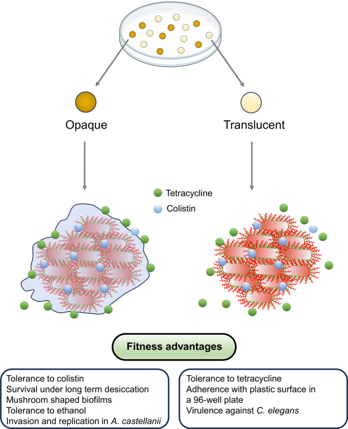
Carbapenem-resistant Acinetobacter baumannii causes one of the most difficult-to-treat nosocomial infections. Polycationic drugs like polymyxin B or colistin and tetracycline drugs such as doxycycline or minocycline are commonly used to treat infections caused by carbapenem-resistant A. baumannii. Here, we show that a subpopulation of cells associated with the opaque/translucent colony phase variation by A. baumannii AB5075 displays differential tolerance to subinhibitory concentrations of colistin and tetracycline. Using a variety of microscopic techniques, we demonstrate that extracellular polysaccharide moieties mediate colistin tolerance to opaque A. baumannii at single-cell level and that mushroom-shaped biofilm structures protect opaque bacteria at the community level. The colony switch phenotype is found to alter several traits of A. baumannii, including long-term survival under desiccation, tolerance to ethanol, competition with Escherichia coli, and intracellular survival in the environmental model host Acanthamoeba castellanii. Additionally, our findings suggest that extracellular DNA associated with membrane vesicles can promote colony switching in a DNA recombinase-dependent manner.IMPORTANCEAs a WHO top-priority drug-resistant microbe, Acinetobacter baumannii significantly contributes to hospital-associated infections worldwide. One particularly intriguing aspect is its ability to reversibly switch its colony morphotype on agar plates, which has been remarkably underexplored. In this study, we employed various microscopic techniques and phenotypic assays to investigate the colony phase variation switch under different clinically and environmentally relevant conditions. Our findings reveal that the presence of a poly N-acetylglucosamine-positive extracellular matrix layer contributes to the protection of bacteria from the bactericidal effects of colistin. Furthermore, we provide intriguing insights into the multicellular lifestyle of A. baumannii, specifically in the context of colony switch variation within its predatory host, Acanthamoeba castellanii.

Keywords: colisitin; opaque colony; translucent colony.


Conflict of interest statement

The authors declare no conflict of interest.

Figures

Fig 1
Fig 1
Opaque colonies express extracellular polysaccharide moieties. (A) Left panels: Scanning electron microscopy visualization of bacterial cells from opaque and translucent colonies of A. baumannii AB5075. Right panels: Higher magnification images to illustrate the presence of an additional layer of extracellular material in case of opaque colonies. (B) Transmission electron microscopy of individual cells from opaque and translucent colonies of A. baumannii AB5075. (C) Representative confocal laser scanning microscopy images of WGA-labeled opaque and translucent colonies of A. baumannii AB5075. Scale bars = 5 µm. (D) Fluorescence quantification of samples from pair-wise opaque and translucent variants of different A. baumannii clinical isolates and A. haemolyticus upon labeling with Aelxa647-WGA.
Fig 2
Fig 2
WGA-binding extracellular moieties protect opaque A. baumanni from colistin. (A) Confocal laser microscopy images of opaque and translucent variants of A. baumannii AB5075 grown in an 18-well glass chamber upon the treatment of colistin. Propidium iodide was used to differentiate dead and live bacteria upon the treatment with colistin. Red: PI-positive cells (dead cells), Blue: WGA binding representing the presence of extracellular polysaccharide moieties. Scale bars = 5 µm. (B) Left panel: Minimum inhibitory concentration of colistin in opaque and translucent colony variants of A. baumannii strain AB5075 as determined by microbroth dilution method. Right panel: Growth curves of opaque and translucent A. baumannii AB5075 grown in a microtiter plate in LB medium with subinhibitory concentration of colistin at 37°C using the built-in temperature control mode of the Spark multimode plate reader (Tecan). Y-axis represents the optical density (OD600) measured with an interval of 20 min. The experiment was done in triplicate, and the curves were drawn using the mean OD600 values. (C) Left panel: Minimum inhibitory concentration of tetracycline in opaque and translucent colony variants of A. baumannii AB5075 as determined by microbroth dilution method. Right panel: Growth curves of opaque and translucent A. baumannii AB5075 grown in a microtiter plate in LB medium with subinhibitory concentration of tetracycline at 37°C using the built-in temperature control mode of the Spark multimode plate reader (Tecan). Y-axis represents the optical density (OD600) measured with an interval of 20 min. The experiment was done in triplicate, and the curves were drawn using the mean OD600 values.
Fig 3
Fig 3
3D biofilm structures formed by opaque and translucent A. baumannii AB5075. (A) Scanning electron microscopy visualization of bacteria from A. baumannii AB5075 grown in the pellicle formed by the opaque and translucent colony morphotype, respectively. (B) 3D live cell confocal laser microscopy images of biofilms formed by A. baumannii AB5075 opaque and translucent variants expressing green fluorescent protein within static flow cells after 72 h of incubation at 30°C. White arrowhead indicates dense patches of biofilm in opaque variant of A. baumannii. (C) Live cell confocal microscopy image of opaque and translucent variants of A. baumannii AB5075 expressing green fluorescent protein after growth in static flow cells and upon 30 or 60 min treatment with 1 µg/mL colistin. Propidium iodide was used to differentiate dead and live bacteria upon the treatment with colistin. Live cells are shown in green and dead cells are shown in red. Dense patches of biofilm in opaque colonies (as indicated by a white arrowhead) seemed protected from the bactericidal effect of colistin. Scale bars = 5 µm.
Fig 4
Fig 4
Comparative analysis of opaque and translucent variants of A. baumannii for survival under various stress-inducing conditions. (A) The survival curves illustrate the number of bacteria that remained viable in samples representing opaque and translucent variants of clinical isolates under desiccation for the period of up to 150 days. (B) Ethanol tolerance test estimating the survival of opaque and translucent variants of A. baumannii isolates upon the treatment with 5% ethanol in PBS at room temperature for 4 h. (C) Growth curves of opaque and translucent A. baumannii AB5075 grown in a microtiter plate in LB medium supplemented with 5% or 10% ethanol. The experiment was performed at 37°C using the built-in temperature control mode of the Spark multimode plate reader (Tecan). The Y-axis represents the optical density (OD600) measured with an interval of 20 min for 16 h. The curves represent the mean OD600 values from experiment done in triplicate. (D) Photographic images of the mixed colonies of A. baumannii and E. coli on LB agar plates supplemented with carbenicillin, IPTG, and X-gal. White spots represent A. baumannii and blue spots represent E. coli MC 20; Left panel: initial inoculum consists of 106 bacteria (1:1 ratio of A. baumannii and E. coli); Right panel: initial inoculum consists of 103 bacteria (1:1 ratio of A. baumannii and E. coli). (E) A bar chart diagram compares the number of E. coli MC 20 and A. baumannii variants upon growing together on agar plates.
Fig 5
Fig 5
Interaction of A. baumannii AB5075 with its environmental host A. castellanii. (A) Confocal microscopic visualization of Acanthamoeba castellanii harboring intracellular A. baumannii 48 h after infection. The white arrowhead indicates intracellular localization of EGFP-A. baumannii (green). A. castellanii was stained with actin marker, Phalloidin594 (red). Scale bars = 5 µm. (B) Confocal microscopic visualization of phalloidin labeled (Actin) A. castellanii harboring intracellular opaque A. baumannii to illustrate localization of intracellular dividing bacteria within a distinct vacuole. Scale bars = 10 µm. (C) Percent recovery of A. baumannii AB5075 variants after mixed infection of opaque and translucent A. baumannii with 1:1 ratio.
Fig 6
Fig 6
Effect of outer membrane vesicles and OMVs-bound DNA on colony switch phenotype. (A) Atomic force microscopic visualization of A. baumannii AB5075 opaque (left panel) and translucent (right panel) variants along with secreted vesicles. Bar chart diagrams to illustrate switching frequencies of A. baumannii AB5075 from opaque to translucent (B) and translucent to opaque (C) upon supplementation of MVs prep at a concentration of 100 mg/mL of total protein contents. (D) Column bar diagram to illustrate the effect of MVs treated with DNAase I and proteinase K (1U each per 100 mg of total protein at 37°C) on the colony switching phenotype. For enzymatic treatment, OMVs were incubated with DNAse 1 (1U/100 mg of MVs protein content) for 30 min at 37°C. Subsequently, proteinase K (1U/100 mg of MVs protein content were added). The suspension was again incubated for 30 min at 37°C. The enzymes were inactivated by incubation of the suspension at 65°C for 10 min. The values shown in Y-axis are relative colony count as compared to average number of translucent colonies in the absence of any treatment. CM; sterile conditioned medium obtained after filtering the overnight culture of A. baumannii AB5075OP. (E) Column bar diagram to compare frequency of opaque to translucent switch in transposon insertion mutants of A. baumannii AB5075 recA::Tn and uvrD::Tn with wild type (WT). (F) Column bar diagram to compare frequency of opaque to translucent switch in A. baumannii AB5075 upon overexpression of RecAB from the plasmid. (G) A representative image of A. baumannii AB5075 transformants of pAT04 (pMBB67EH-RecET: Tet) plasmid; Left panel: opaque colonies with overproduction of RecET harbor multiple translucent sectors as shown with rectangle. Right panel: Bacterial colonies upon the plating of 1/105 dilution of the colony shown in left panel. (H) Bar chart diagram to show switching frequency upon over production of RecET in tetracycline plates as compared to vector control.
Fig 7
Fig 7
Schematic diagram to summarize fitness advantages of opaque and translucent colony morphotypes of A. baumannii. The production of extracellular sheet protects opaque variant from suboptimal concentration of colistin. The fitness advantages here summarized also include findings of our previous study (7).

Similar articles

Cited by

References

    1. Giamarellou H, Antoniadou A, Kanellakopoulou K. 2008. Acinetobacter baumannii: a universal threat to public health Int J Antimicrob Agents 32:106–119. doi:10.1016/j.ijantimicag.2008.02.013 - DOI - PubMed
    1. Ferguson WW, Roberts LF. 1950. A bacteriological and serological study of organism B5W (bacterium antiratum). J Bacteriol 59:171–183. doi:10.1128/jb.59.2.171-183.1950 - DOI - PMC - PubMed
    1. Chin CY, Tipton KA, Farokhyfar M, Burd EM, Weiss DS, Rather PN. 2018. A high-frequency phenotypic switch links bacterial virulence and environmental survival in Acinetobacter baumannii. Nat Microbiol 3:563–569. doi:10.1038/s41564-018-0151-5 - DOI - PMC - PubMed
    1. Tierney ARP, Chin CY, Weiss DS, Rather PN. 2021. A Lysr-type transcriptional regulator controls multiple phenotypes in Acinetobacter baumannii Front Cell Infect Microbiol 11:778331. doi:10.3389/fcimb.2021.778331 - DOI - PMC - PubMed
    1. Tipton KA, Dimitrova D, Rather PN. 2015. Phase-variable control of multiple phenotypes in Acinetobacter baumannii strain AB5075. J Bacteriol 197:2593–2599. doi:10.1128/JB.00188-15 - DOI - PMC - PubMed

LinkOut - more resources