. 2024 Mar;242(3):543-557.
doi: 10.1007/s00221-023-06763-x. Epub 2024 Jan 11.

Vivaria housing conditions expose sex differences in brain oxidation, microglial activation, and immune system states in aged hAPOE4 mice

Affiliations

Vivaria housing conditions expose sex differences in brain oxidation, microglial activation, and immune system states in aged hAPOE4 mice

E M Reyes-Reyes et al. Exp Brain Res. 2024 Mar.

Abstract

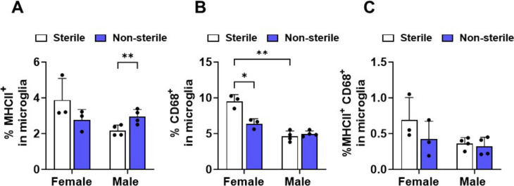
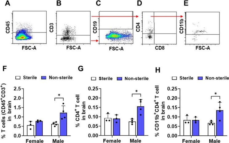
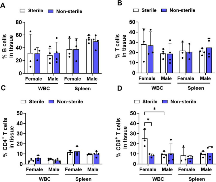
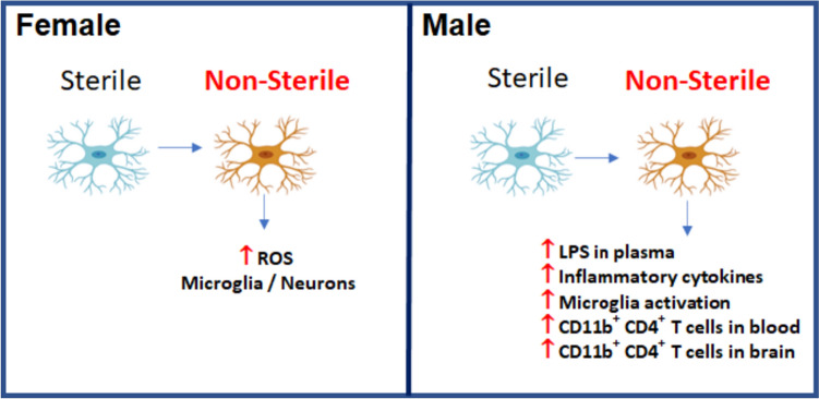
Apolipoprotein E ε4 allele (APOE4) is the predominant genetic risk factor for late-onset Alzheimer's disease (AD). APOE4 mouse models have provided advances in the understanding of disease pathogenesis, but unaccounted variables like rodent housing status may hinder translational outcomes. Non-sterile aspects like food and bedding can be major sources of changes in rodent microflora. Alterations in intestinal microbial ecology can cause mucosal barrier impairment and increase pro-inflammatory signals. The present study examined the role of sterile and non-sterile food and housing on redox indicators and the immune status of humanized-APOE4 knock-in mice (hAPOe4). hAPOE4 mice were housed under sterile conditions until 22 months of age, followed by the transfer of a cohort of mice to non-sterile housing for 2 months. At 24 months of age, the redox/immunologic status was evaluated by flow cytometry/ELISA. hAPOE4 females housed under non-sterile conditions exhibited: (1) higher neuronal and microglial oxygen radical production and (2) lower CD68+ microglia (brain) and CD8+ T cells (periphery) compared to sterile-housed mice. In contrast, hAPOE4 males in non-sterile housing exhibited: (1) higher MHCII+ microglia and CD11b+CD4+ T cells (brain) and (2) higher CD11b+CD4+ T cells and levels of lipopolysaccharide-binding protein and inflammatory cytokines in the periphery relative to sterile-housed mice. This study demonstrated that sterile vs. non-sterile housing conditions are associated with the activation of redox and immune responses in the brain and periphery in a sex-dependent manner. Therefore, housing status may contribute to variable outcomes in both the brain and periphery.

Keywords: Alzheimer’s disease; Apolipoprotein; Housing and food sterility; Immune status; Sex differences.


Conflict of interest statement

The authors declare that they have no competing interests that could have influenced the work presented in this manuscript. Financial interests, professional relationships, or personal connections that might have a potential conflict of interest with the research findings are absent.

Figures

Fig. 1
Fig. 1
Oxidant production is increased in non-sterile conditions in hAPOE4 female, but not male, brain cells. Brain cells were dissociated from whole brain samples, stained for type-specific markers, and analyzed by flow cytometry. Panels (A–H) are representative graphs illustrating the brain cell gating strategy. A Brain cells were selected by their SSC and FSC characteristics. B Dead brain cells and C cell doublets were discriminated by their DAPI staining and FSC-A and FSC-H characteristics. Single brain cells were identified by gating expression of specific cell type markers: D Microglia expressing CD11b high and CD45 intermediate, E Astrocytes expressing ACSA-2+/CD45/CD11b, neurons expressing CD31/O4/ACSA-2/CD45/CD11b, F oligodendrocyte expressing O4+/CD31/ACSA-2/CD45/CD11b and endothelial cells by expression of CD31+/O4/ACSA-2/CD45/CD11b. G Each brain cell type was used for gating mitochondrial oxidative stress (MitoSox+). H Microglial cells were gated for expression of MHC-II and CD68. Quantification of mitochondrial oxidative stress in microglia (I) and neurons (J). Data are represented as mean ± SD. p values were calculated using two-way ANOVA (Bonferroni adjusted p-values are shown in Supplementary Table 2; *p ≤ 0.05, **p ≤ 0.01)
Fig. 2
Fig. 2
Microglia activation is affected by housing and food status. Quantification of expression of microglial activation markers A MHCII+, B CD68+, and C MHCII+ CD68+. Data are represented as mean ± SD. p values calculated using two-way ANOVA (Bonferroni adjusted p-values are shown in Supplementary Table 2; *p ≤ 0.05, **p ≤ 0.01, and ***p ≤ 0.001)
Fig. 3
Fig. 3
Non-sterile environments increase the infiltration of T cells into the male, but not female brain. Male and female APOE4 mouse brains were dissociated and assessed using a single-cell suspension. Brain cells were stained for immune cell markers and analyzed by flow cytometry. A After discriminating dead cells and doublets, immune cells were gated by expressing CD45+. Immune cells were classified as B T cells (CD45+ CD3+), C B cells (CD19+/CD45+/CD3) and D helper T cells (CD4+/CD3+/CD45+) and cytotoxic T cells (CD8+/CD3+/CD45+). E Helper T cells were gated for expression of CD11b. Quantification of F total T cells, G helper T cells and H CD11b+ helper T cells in the brain. Data are represented as mean ± SD. p values were calculated using two-way ANOVA (Bonferroni adjusted p-values are shown in Supplementary Table 2; *p ≤ 0.05, and **p ≤ 0.01)
Fig. 4
Fig. 4
Peripheral adaptive immune cells are affected by sterile and non-sterile food and housing conditions. White blood cells (WBC) and splenic cells were isolated and stained immune cell markers and analyzed by flow cytometry. A Quantification of % B cells (CD19+/CD45+/CD3), B T cell (CD3+/CD45+), C helper T cell (CD4+/CD3+/CD45+) and D cytotoxic T cells (CD8+/CD3+/CD45+) in spleen and WBC. Data are represented as mean ± SD. p values were calculated using two-way ANOVA (Bonferroni adjusted p-values are shown in Supplementary Table 2; *p ≤ 0.05, **p ≤ 0.01, ***p ≤ 0.001)
Fig. 5
Fig. 5
Sterile and non-sterile food and housing effects on the expression of T cell activation and inflammatory markers. Splenic cells and WBCs were stained for B cell and T cell markers (CD3+, CD4+, CD8+), the T cell activation marker CD69+, and inflammation marker CD11b+ and analyzed by flow cytometry. CD69+ was measured in both A CD4+ and B CD8+ T cells in the blood and in the spleen (C, D). CD11b+ was measured in C CD4+ and D CD8+ T cells and E B cells in the blood and in the spleen. Data are represented as mean ± SD. p values were calculated using two-way ANOVA (Bonferroni adjusted p-values are shown in Supplementary Table 2; *p ≤ 0.05 and **p ≤ 0.01)
Fig. 6
Fig. 6
Non-sterile food and housing increase the levels of LPS-binding protein and inflammatory cytokines in the plasma of APOE4 males. The concentration of circulating A LPS-binding protein (LBP), B TNF-α, C IL-1β, D KC/GRO, E IL-6, and F INF-γ- were measured from plasma collected at necropsy. Data are represented as mean ± SD. p values were calculated using two-way ANOVA (Bonferroni adjusted p-values are shown in Supplementary Table 2; *p ≤ 0.05 and **p ≤ 0.01)
Fig. 7
Fig. 7
Aged hAPOE4 males are more susceptible to housing and food status than aged hAPOE4 females. hAPOE4 females and males responded differently to sterile and non-sterile housing and food status. Female hAPOE4 mice moved to non-sterile conditions increase in MitoSox + microglia and neurons and did not exhibit an inflammatory response to non-sterile food and housing conditions. However, male hAPOE4 mice exposed to non-sterile food and housing exhibited an increase in the levels of LPS and inflammatory cytokines in the circulatory system, activated microglia, infiltration of T cells into the brain compared to those in sterile food and housing conditions

References

    1. Altmann A, Tian L, Henderson VW, Greicius MD, Alzheimer's Disease Neuroimaging Initiative I (2014) Sex modifies the APOE-related risk of developing Alzheimer disease. Ann Neurol 75:563–573. 10.1002/ana.24135 - PMC - PubMed
    1. Alzheimer’s Association Report 2021 Alzheimer's disease facts and figures. Alzheimers Dement. 2021;17:327–406. doi: 10.1002/alz.12328. - DOI - PubMed
    1. Alzheimer’s Association Report AAIC 2021: The promise of Alzheimer's research. Alzheimers Dement. 2021;17:1868–1871. doi: 10.1002/alz.12500. - DOI - PubMed
    1. Arnold M, Nho K, Kueider-Paisley A, et al. Sex and APOE epsilon4 genotype modify the Alzheimer's disease serum metabolome. Nat Commun. 2020;11:1148. doi: 10.1038/s41467-020-14959-w. - DOI - PMC - PubMed
    1. Balu D, Karstens AJ, Loukenas E, Maldonado Weng J, York JM, Valencia-Olvera AC, LaDu MJ. The role of APOE in transgenic mouse models of AD. Neurosci Lett. 2019;707:134285. doi: 10.1016/j.neulet.2019.134285. - DOI - PMC - PubMed

Substances