. 2024 May;40(5):1377-1388.
doi: 10.1007/s00381-024-06275-1. Epub 2024 Jan 11.

CSF and venous blood flow from childhood to adulthood studied by real-time phase-contrast MRI

Affiliations

CSF and venous blood flow from childhood to adulthood studied by real-time phase-contrast MRI

Prativa Sahoo et al. Childs Nerv Syst. 2024 May.

Abstract

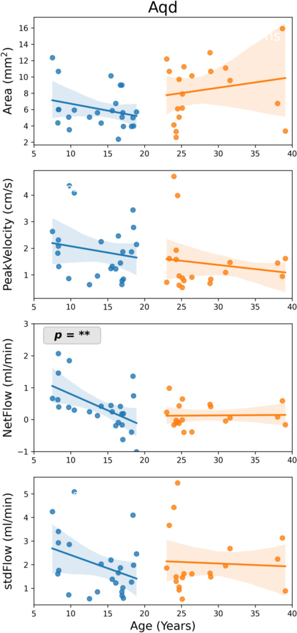
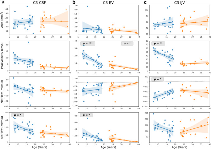
Purpose: In vivo measurements of CSF and venous flow using real-time phase-contrast (RT-PC) MRI facilitate new insights into the dynamics and physiology of both fluid systems. In clinical practice, however, use of RT-PC MRI is still limited. Because many forms of hydrocephalus manifest in infancy and childhood, it is a prerequisite to investigate normal flow parameters during this period to assess pathologies of CSF circulation. This study aims to establish reference values of CSF and venous flow in healthy subjects using RT-PC MRI and to determine their age dependency.

Methods: RT-PC MRI was performed in 44 healthy volunteers (20 females, age 5-40 years). CSF flow was quantified at the aqueduct (Aqd), cervical (C3) and lumbar (L3) spinal levels. Venous flow measurements comprised epidural veins, internal jugular veins and inferior vena cava. Parameters analyzed were peak velocity, net flow, pulsatility, and area of region of interest (ROI).

Statistical tests: linear regression, student's t-test and analysis of variance (ANOVA).

Results: In adults volunteers, no significant changes in flow parameters were observed. In contrast, pediatric subjects exhibited a significant age-dependent decrease of CSF net flow and pulsatility in Aqd, C3 and L3. Several venous flow parameters decreased significantly over age at C3 and changed more variably at L3.

Conclusion: Flow parameters varies depending on anatomical location and age. We established changes of brain and spinal fluid dynamics over an age range from 5-40 years. The application of RT-PC MRI in clinical care may improve our understanding of CSF flow pathology in individual patients.

Keywords: Age-related changes of flow; CSF dynamics; Childhood hydrocephalus; Real-time phase-contrast MRI; Venous flow.


Conflict of interest statement

The other authors declare no competing financial interest.

Figures

Fig. 1
Fig. 1
ROIs for CSF and venous flow analyses. a The sagittal image indicates the slice locations chosen for Aqd, spinal level C3 and L3. ROIs are shown on axial magnitude images: b Aqd, c C3 CSF (yellow) and C3 EV (blue), d C3 IJV, e L3 CSF (yellow) and L3 EV (blue), and f L3 IVC. ROI = region of interest, Aqd = aqueduct, C3 = cervical spinal level 3, L3 = lumbar spinal level 3, EV = epidural veins, IJV = internal jugular vein, IVC = inferior vena cava
Fig. 2
Fig. 2
Linear regression between flow parameters and age at the aqueduct. Parameters are (from top to bottom): ROI Area (mm2), peakVelocity (cm/s), netFlow (ml/min), and stdFlow (ml/min). Stars indicate a slope significantly different from zero, i.e. p < 0.05. Shaded regions represent 95% confidence interval. Aqd = aqueduct, stdFlow = standard deviation over flow time series
Fig. 3
Fig. 3
Linear regression between flow parameters and age at cervical spinal level C3. Parameters are (from top to bottom): ROI Area (mm2), peakVelocity (cm/s), netFlow (ml/min), and stdFlow (ml/min). ROIs from left to right C3 CSF (a), C3 EV (b), C3 IJV (c). CSF flow is shown in left column, venous flow in middle and right columns. Stars indicate a slope significantly different from zero, i.e. p < 0.05. Shaded regions represent 95% confidence interval. C3 = cervical spinal level 3, stdFlow = standard deviation over flow time series, EV = epidural veins, IJV = internal jugular vein
Fig. 4
Fig. 4
Linear regression between flow parameters and age at lumbar spinal level L3. Parameters are (from top to bottom): ROI Area (mm2), peakVelocity (cm/s), netFlow (ml/min), and stdFlow (ml/min). Stars indicate a slope significantly different from zero, i.e. p < 0.05. Shaded regions represent 95% confidence interval. CSF flow is shown in left column, venous flow in middle and right column. L3 = lumbar spinal level 3, stdFlow = standard deviation over flow time series, EV = epidural veins, IVC = inferior vena cava

References

    1. Mohammad SA, Osman NM, Ahmed KA. The value of CSF flow studies in the management of CSF disorders in children: a pictorial review. Insights Imaging. 2019;10:3. doi: 10.1186/s13244-019-0686-x. - DOI - PMC - PubMed
    1. Plata KS, Cruz G, Lezcano H. Cine phase-contrast magnetic resonance imaging evaluation of cerebrospinal fluid flow dynamics in healthy pediatric subjects. Radiol Bras. 2022;55:225–230. doi: 10.1590/0100-3984.2021.0120. - DOI - PMC - PubMed
    1. Yamada S, Tsuchiya K, Bradley WG, et al. Current and emerging MR imaging techniques for the diagnosis and management of CSF flow disorders: a review of phase-contrast and time-spatial labeling inversion pulse. AJNR Am J Neuroradiol. 2015;36:623–630. doi: 10.3174/ajnr.A4030. - DOI - PMC - PubMed
    1. Naidich TP, Altman NR, Gonzalez-Arias SM. Phase contrast cine magnetic resonance imaging: normal cerebrospinal fluid oscillation and applications to hydrocephalus. Neurosurg Clin N Am. 1993;4:677–705. doi: 10.1016/S1042-3680(18)30559-X. - DOI - PubMed
    1. Sohns JM, Kowallick JT, Joseph AA, et al. Peak flow velocities in the ascending aorta-real-time phase-contrast magnetic resonance imaging vs. cine magnetic resonance imaging and echocardiography. Quant Imaging Med Surg. 2015;5:685–690. doi: 10.3978/j.issn.2223-4292.2015.08.08. - DOI - PMC - PubMed