Review
. 2024 Jul 1;104(3):881-929.
doi: 10.1152/physrev.00024.2023. Epub 2024 Jan 11.

The anterior chamber of the eye technology and its anatomical, optical, and immunological bases

Affiliations
Review

The anterior chamber of the eye technology and its anatomical, optical, and immunological bases

Shao-Nian Yang et al. Physiol Rev. .

Abstract

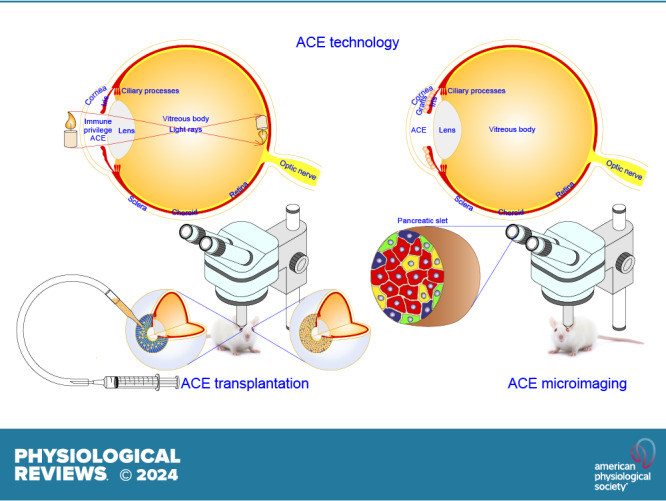
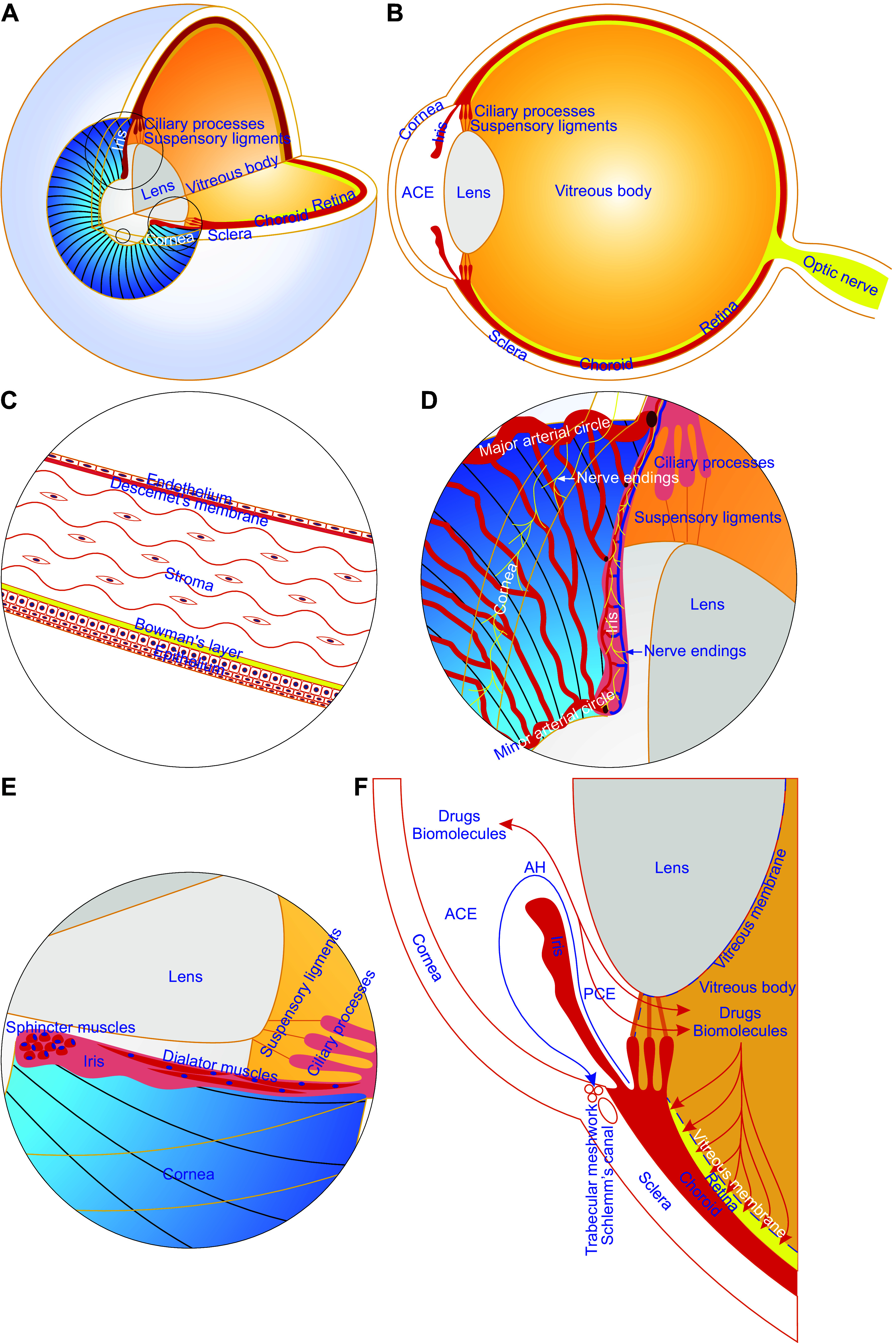
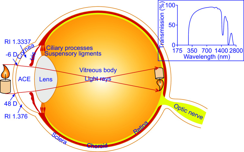
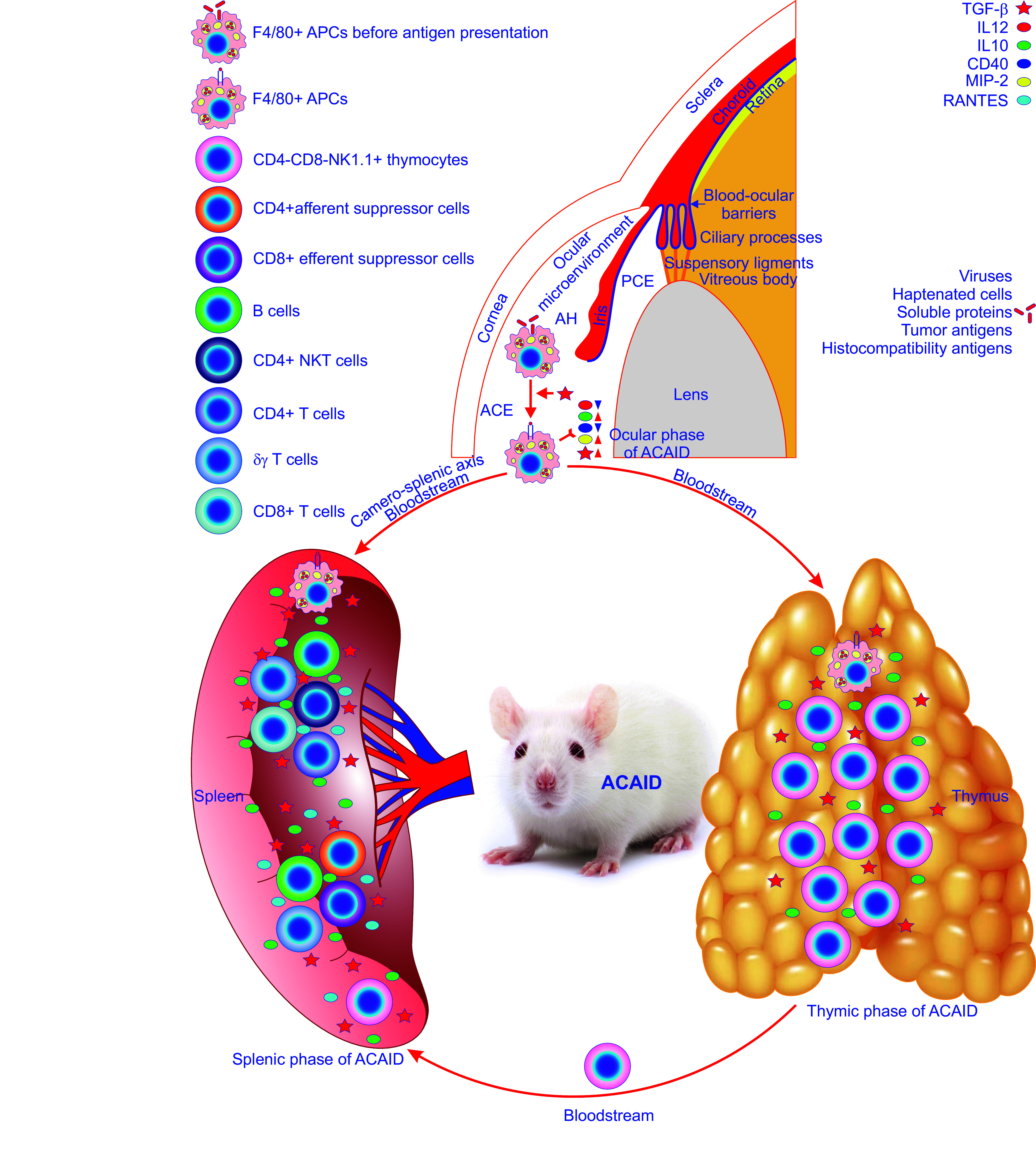
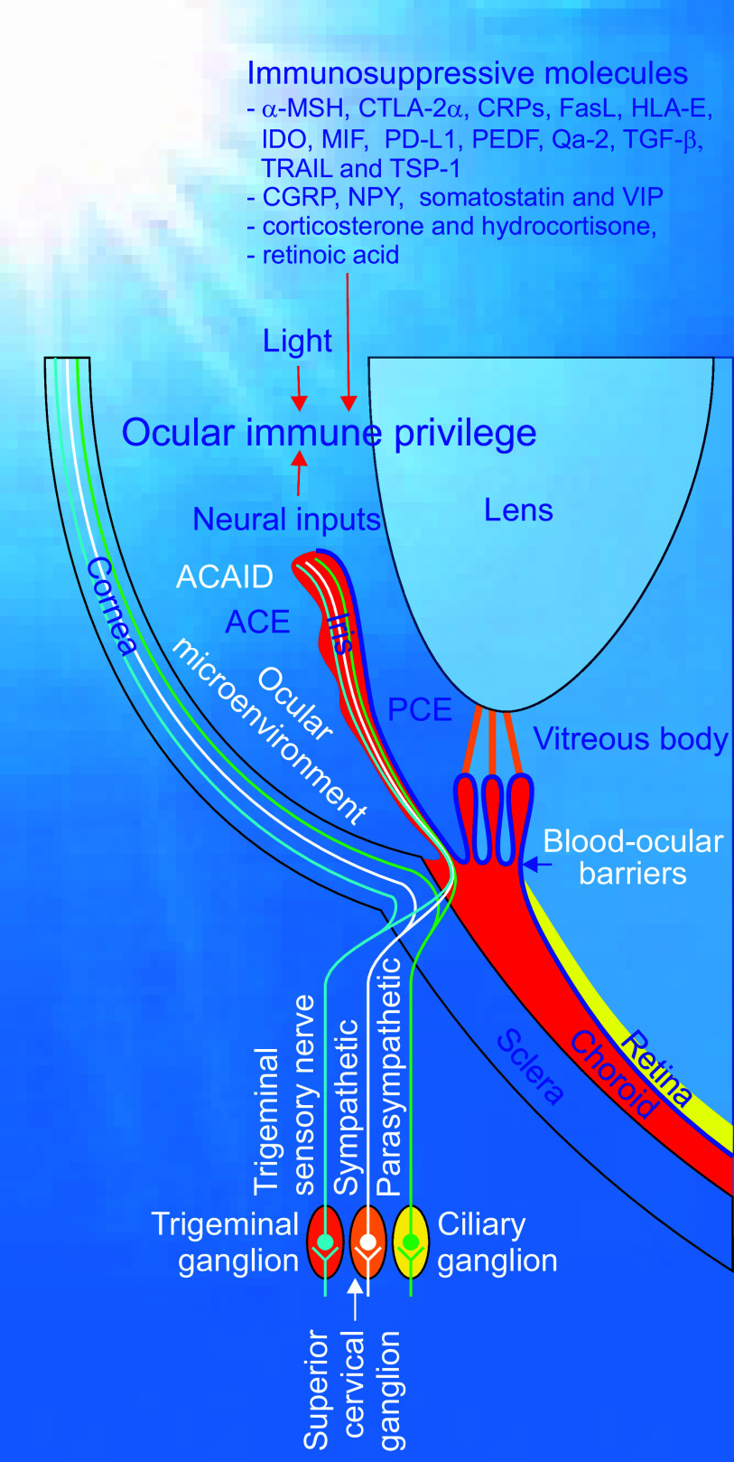
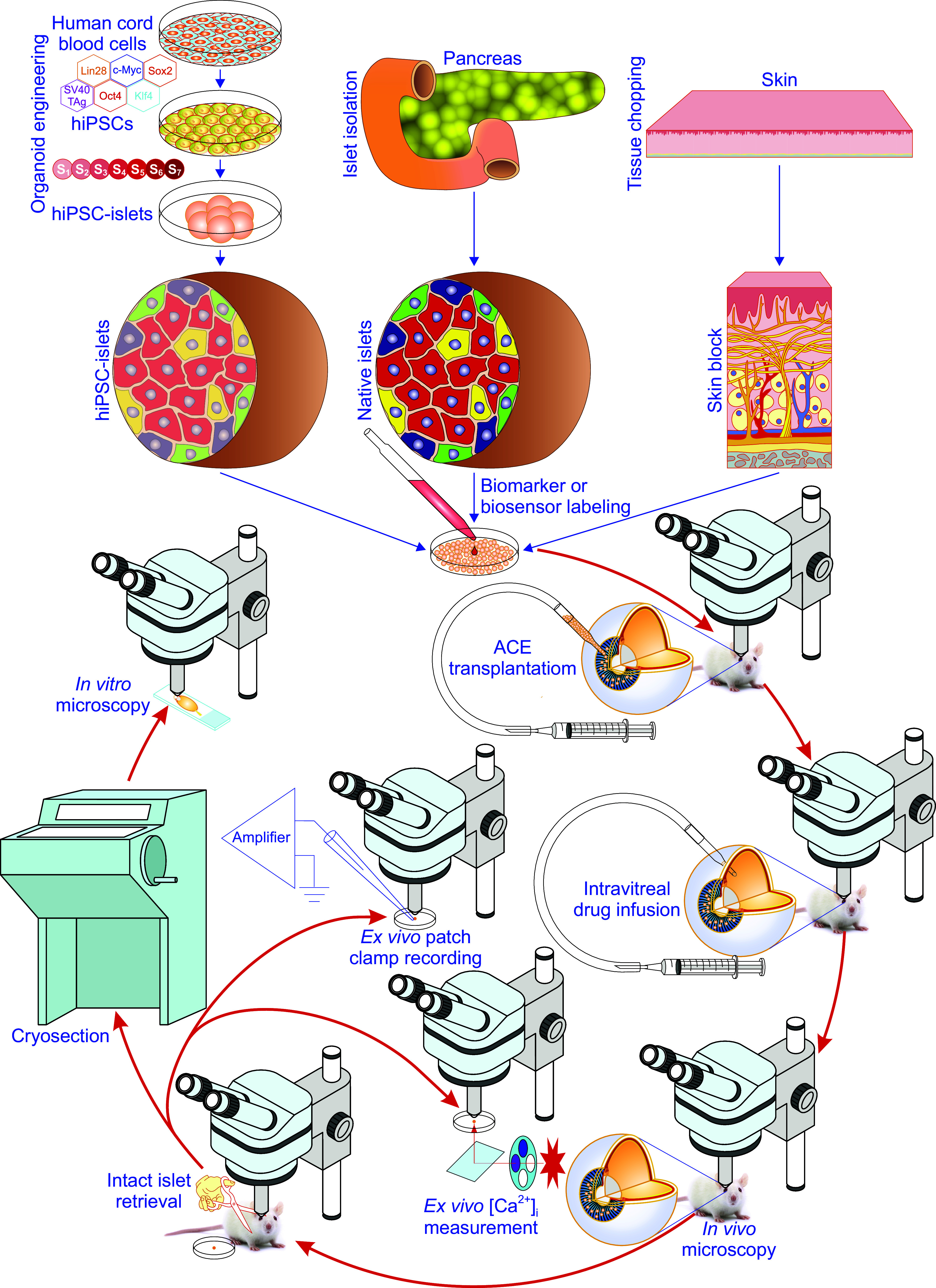
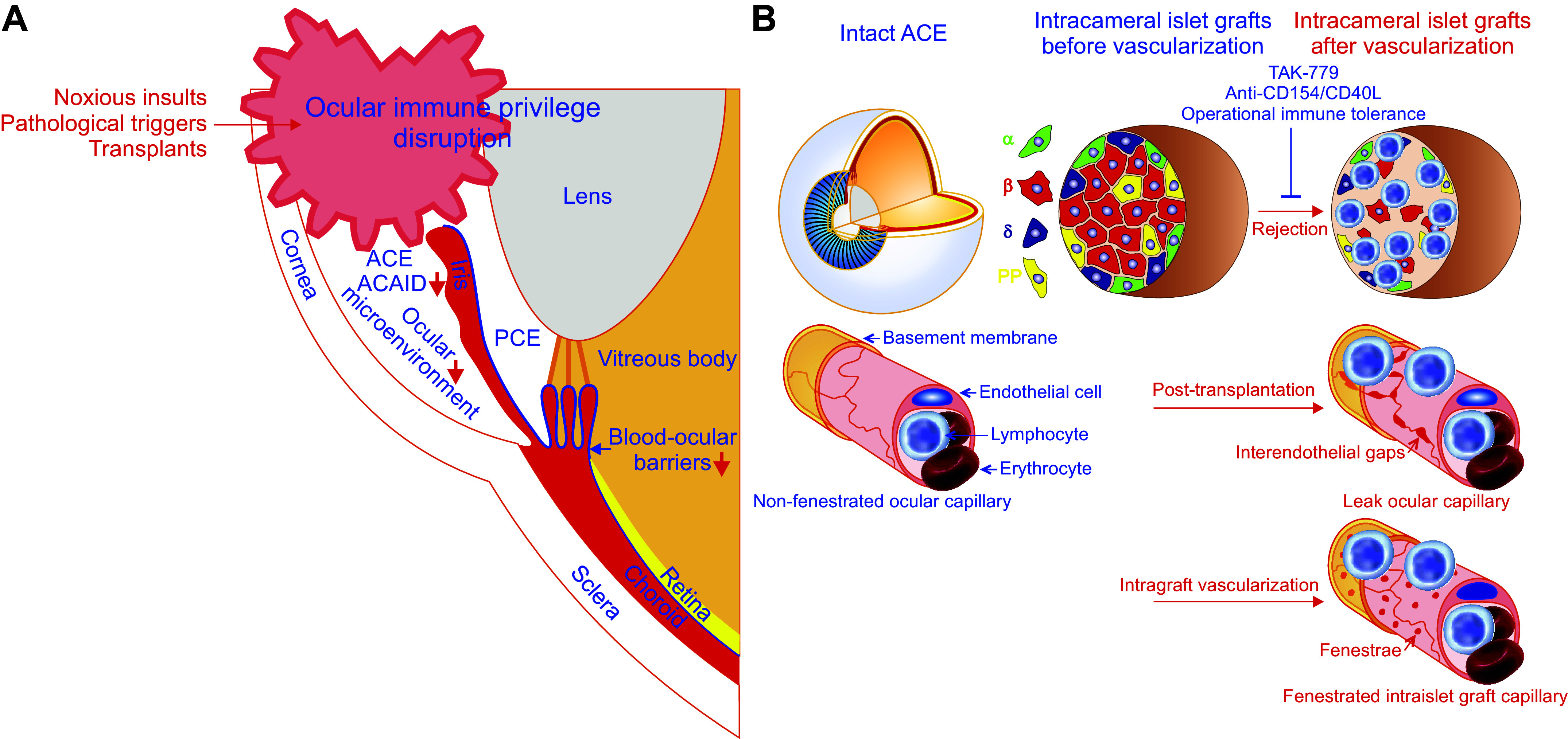
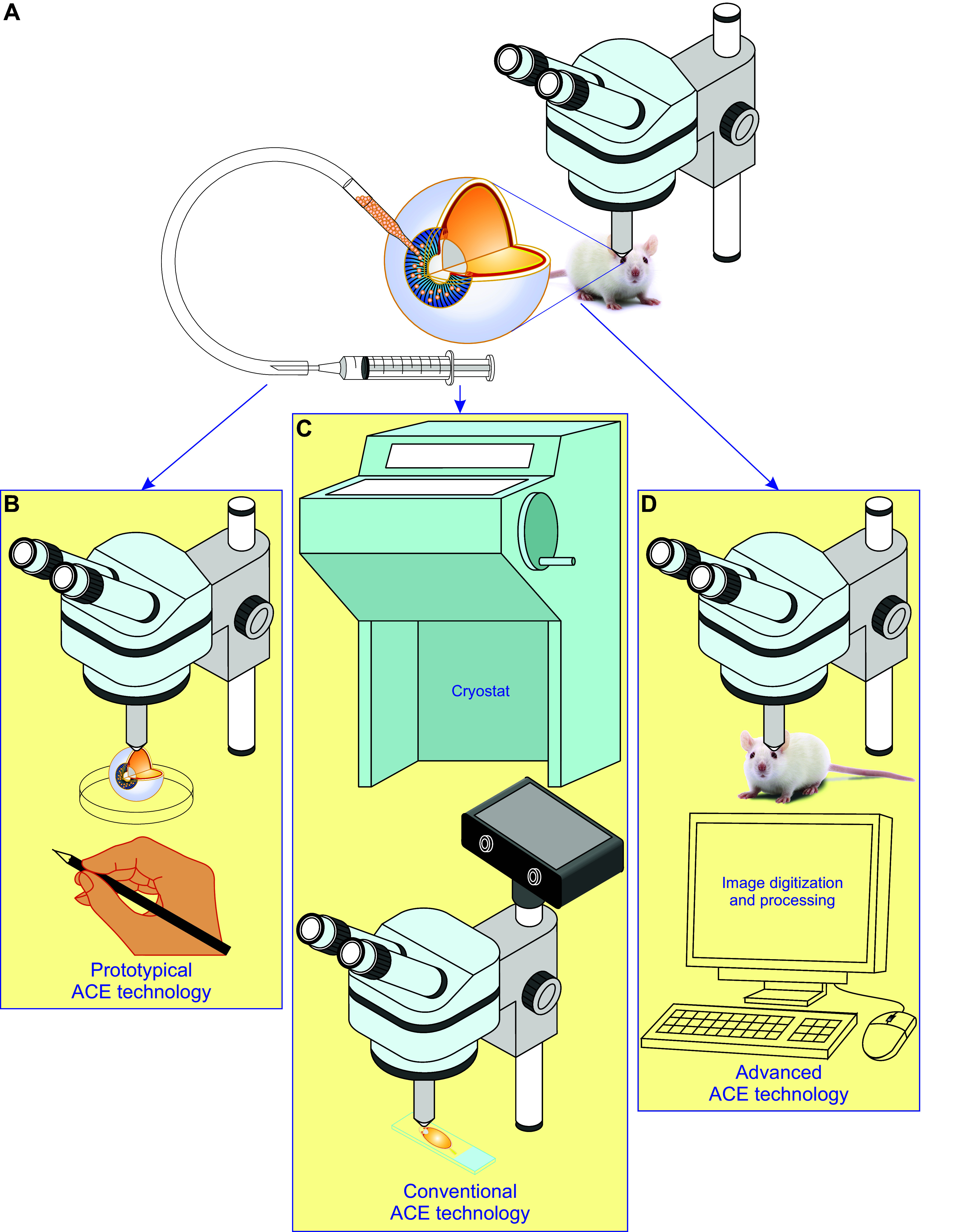
The anterior chamber of the eye (ACE) is distinct in its anatomy, optics, and immunology. This guarantees that the eye perceives visual information in the context of physiology even when encountering adverse incidents like inflammation. In addition, this endows the ACE with the special nursery bed iris enriched in vasculatures and nerves. The ACE constitutes a confined space enclosing an oxygen/nutrient-rich, immune-privileged, and less stressful milieu as well as an optically transparent medium. Therefore, aside from visual perception, the ACE unexpectedly serves as an excellent transplantation site for different body parts and a unique platform for noninvasive, longitudinal, and intravital microimaging of different grafts. On the basis of these merits, the ACE technology has evolved from the prototypical through the conventional to the advanced version. Studies using this technology as a versatile biomedical research platform have led to a diverse range of basic knowledge and in-depth understanding of a variety of cells, tissues, and organs as well as artificial biomaterials, pharmaceuticals, and abiotic substances. Remarkably, the technology turns in vivo dynamic imaging of the morphological characteristics, organotypic features, developmental fates, and specific functions of intracameral grafts into reality under physiological and pathological conditions. Here we review the anatomical, optical, and immunological bases as well as technical details of the ACE technology. Moreover, we discuss major achievements obtained and potential prospective avenues for this technology.

Keywords: in vivo microimaging; innervation; the anterior chamber of the eye; transplantation; vascularization.


Conflict of interest statement

S.-N. Yang is a consultant to Biocrine AB. P.-O. Berggren is the cofounder and CEO of BioCrine AB.

Figures

None
Graphical abstract
FIGURE 1.
FIGURE 1.
Anatomical features of the anterior chamber of the eye (ACE). The ACE is built up mainly of the cornea, iris, and aqueous humor (AH). The cornea is avascular and consists of a thin nonkeratinized epithelium, acellular Bowman’s layer, stroma with highly organized collagen fibrils and very few keratocytes, cell-free matrix-formed Descemet’s membrane, and endothelial cell monolayer. Hence, the cornea is characterized as a transparent natural body window that allows light to reach the retina and gives rise to a key prerequisite for noninvasive, longitudinal, and intravital microimaging of intracameral grafts. The iris is richly vascularized and innervated. It can provide a major engraftment area for intracameral grafts and predominately contributes to intracameral graft vascularization and innervation, thereby acting as a favorable graft survival substratum. The AH is a water-like liquid, produced by the ciliary processes, flows into the posterior chamber of the eye and then the ACE through the pupil and eventually drains sequentially into trabecular meshworks, Schlemm’s canal, and episcleral veins. The AH supplies oxygen, nutrients, and other survival factors to and removes metabolic wastes from intracameral grafts. Of note, the vitreous membrane is porous and allows diffusion of intraocular drugs or biomolecules between the anterior/posterior chambers, vitreous body, and retinal compartment. A and B: three-fourths 3-dimensional and one-half sagittal views of the eyeball showing the structure of the ACE. C: partially enlarged view of the small circle area in A showing the 5-layer architecture of the cornea. D and E: partially enlarged view of the large and middle circle areas in A showing iridic vasculatures, nerve endings, and smooth muscles. F: sectional view of the partial lower half eyeball illustrating the drainage path for AH and the diffusion paths of intraocular drugs and biomolecules between intraocular compartments. PCE, posterior chamber of the eye.
FIGURE 2.
FIGURE 2.
The optical quality of the anterior chamber of the eye (ACE). The transparency and absorbance of the cornea and aqueous humor (AH) as well as the shape of the former determine the optical quality of the ACE. The cornea refracts light twice when entering and leaving the cornea. Light enters the cornea at the greater angle than it leaves it because the difference in refractive index (RI) between cornea and AH is smaller than that between the air and cornea. The cornea and AH also serve as filters rejecting some of the most damaging ultraviolet rays and getting rid of significant amounts of infrared waves at 1,430 nm and 1,950 nm. D, diopter. Inset is adapted from Ref. , with permission.
FIGURE 3.
FIGURE 3.
The constitution of ocular immune privilege. Ocular immune privilege is attributed to a diverse range of ocular and systemic mechanisms, 3 of which stand out. The first one sequestrates antigens entering the eye due to efficient blood-ocular barriers, including the blood-retinal barrier and the blood-aqueous barrier, and the lack of efferent lymphatics. These 2 barriers are emphasized by a blue line in the eyeball. The second one manifests as active immune suppression by antigens, including viruses, haptenated cells, soluble proteins, tumor antigens, and histocompatibility antigens, invading the anterior chamber of the eye (ACE), i.e., the anterior chamber-associated immune deviation (ACAID). ACAID can be divided into the ocular, thymic, and splenic phases. It starts from the ocular phase where F4/80+ ocular antigen-presenting cells (APCs) capture, process and present invading antigens thereby being primed under regulation of various cytokines and chemokines such as transforming growth factor-β (TGF-β). These primed ocular APCs release less TH1-inducing cytokine IL-12 and CD40 costimulatory molecule and more cytokine IL-10 and become able to produce TGF-β and macrophage inflammatory protein-2 (MIP-2; top). Then, they escape from the ACE into the thymus and spleen through the bloodstream. The latter route is termed the camero-splenic axis. Upon entry into the thymus, the primed ocular APCs induce the thymic phase of ACAID resulting in the generation of CD4-CD8-NK1.1+ thymocytes. Subsequently, these thymocytes flow into the bloodstream and home to the spleen to participate in the splenic phase of ACAID. As soon as the primed F4/80+ ocular APCs reach the spleen, the splenic phase of ACAID commences. In this important phase, F4/80+ APCs, CD4-CD8-NK1.1+ thymocytes, B cells, CD4+ natural killer (NK)T cells, CD4+ T cells, δγ T cells, and CD8+ T cells interact with each other in a milieu enriched in TGF-β, IL12, IL10, CD40, MIP-2, and regulated on activation normal T-expressed and presumably secreted (RANTES). Eventually, this complex immune cell interaction reaches a finale with the production of CD4+ afferent and CD8+ efferent suppressor cells, the former suppressing the induction of DTH responses whereas the latter inhibiting the expression of DTH responses, thereby ultimately resulting in ACAID. The third one is the ocular microenvironment created by ocular cells, such as corneal endothelial cells, iris pigment epithelial cells, ciliary body pigment epithelial cells, and retinal pigment epithelial cells, which secrete immunosuppressive factors and mediate contact-dependent immune suppression. AH, aqueous humor.
FIGURE 4.
FIGURE 4.
The regulation of ocular immune privilege. Ocular immune privilege is tightly regulated by immunosuppressive molecules including cytokines, chemokines, lymphokines, neuropeptides, or other anti-inflammatory species as well as by various signals derived from light exposure and neural inputs from sympathetic, parasympathetic, and sensory nerves to maintain the ocular immunological homeostasis. ACE, anterior chamber of the eye; PCE, posterior chamber of the eye; α-MSH, α-melanocyte stimulating hormone; CGRP, calcitonin gene-related protein; CTLA-2α, cytotoxic T lymphocyte antigen-2α; CRPs, complement regulatory proteins; FasL, Fas ligand; HLA-E, human leukocyte antigen E; IDO, indoleamine dioxygenase; MIF, macrophage migration inhibitory factor; NPY, neuropeptide Y; PD-L1, programmed death-ligand 1; PEDF, pigment epithelium-derived factor; TGF-β, transforming growth factor-β; TRAIL, tumor necrosis factor-related apoptosis-inducing ligand; TSP-1, thrombospondin-1; VIP, vasoactive intestinal peptide.
FIGURE 5.
FIGURE 5.
Methodological details of the anterior chamber of the eye (ACE) technology. Experimentally, the ACE technology can be carried through in the following order (–7, 36, 63). First, different grafts like in vitro-engineered islets from human induced pluripotent stem cells (hiPSCs), isolated native islets from pancreata, and finely chopped skin are prepared and tested for quality in vitro. Second, grafts are genetically labeled with biomarkers or biosensors. This step is only necessary for intravital microimaging. Third, grafts are transplanted into the ACE. After the above 3 steps, there are multiple options including intravital microscopy, intravitreal drug infusion, intact retrieval of intracameral grafts for ex vivo cytoplasmic-free Ca2+ concentration ([Ca2+]i) measurement, ex vivo patch-clamp recording, and histological labeling/in vitro microscopy (–7, 36, 63).
FIGURE 6.
FIGURE 6.
Ocular immune privilege disruption, intracameral alloislet rejection, and tolerance induction. A: the ocular immune privilege can be disrupted by noxious insults, pathological triggers, and transplants, which cause the breakdown of the blood-ocular barrier due to inflammation, ocular neovascularization or transplant revascularization, impair anterior chamber-associated immune deviation (ACAID) induction, and derange ocular microenvironment. B: intracameral alloislet rejection occurs alongside ocular immune privilege disruption induced by islet transplants, which can change nonfenestrated ocular capillaries into leak ones with interendothelial gaps and undergo vascularization characterized by fenestrated intraislet graft capillaries during posttransplantation (29). This alllorejection can be satisfactorily intervened by blocking T-cell chemokine receptors CCR5 and CXCR3 with TAK-779 or the binding of CD154/CD40L to CD40 primarily expressed on activated T cells with anti-CD154/CD40L blocking antibody, the latter reflecting induction of operational immune tolerance (22, 114). ACE, anterior chamber of the eye; PCE, posterior chamber of the eye; PP, pancreatic polypeptide.
FIGURE 7.
FIGURE 7.
Evolution of the anterior chamber of the eye (ACE) technology. This technology is based on anatomical, optical, and immunological features of the ACE and has been developed with the advance of microscopic tools, fluorescent labeling, and image digitization/processing as well as relevant knowledge. A: ACE technology is built up essentially from transplantation of a batch of cells, pieces of tissues and organs, artificial biomaterials, pharmaceuticals, or abiotic substances into the ACE and has evolved into the following three versions. B: prototypical ACE technology, established by van Dooremaal in 1873, simply entails insertion of grafts into the ACE followed by crude observation of intracameral grafts and documentation with freehand drawing (3). C: conventional ACE technology is formed by combining ACE transplantation, a series of in vitro assessments typically by classical histology and microscopy, and objective data analysis and has been widely employed in different fields of biomedicines (38). D: advanced ACE technology is upgraded as a versatile tool with its own uniqueness by advanced microscopy, fluorescent labeling, and image digitization/processing on top of intracameral graft insertion, thus turning noninvasive, longitudinal, and intravital microimaging of different cells, tissues, and organs into reality (–6).
FIGURE 8.
FIGURE 8.
Major achievements by applying the anterior chamber of the eye (ACE) technology. Since its birth, the ACE technology has been successfully applied in various studies on different cells, tissues, and organs as well as artificial biomaterials, pharmaceuticals, and abiotic substances. These studies have brought the following major achievements. In 1873, van Dorremaal (3) found prolonged survival and progressive growth of homologous lip mucosa in the ACE of dogs and rabbits. This finding lays a solid foundation for the development of the concept of ocular immune privilege (3). Seventy-five years later, Medawar (38) immunologically dissected the prolonged survival of intracameral skin allografts of rabbits and coined the concept of ocular immune privilege. In 1972, Gimbrone et al. (372) found that intracameral tumor grafts stopped growing and entered “dormancy” if they were not vascularized and revealed the importance of antineovascular therapy against tumors. Soon after that, Kaplan et al. (149) inoculated allogeneic lymphonoid cells into the rat ACE and proposed “lymphocyte-induced immune deviation” that was subsequently corrected into ACAID. In 2008, Speier et al. (4, 5) succeeded in microimaging intracameral islets in a noninvasive, longitudinal, and intravital manner.
FIGURE 9.
FIGURE 9.
Modes of the anterior chamber of the eye (ACE) technology. The technology can work in the following six different modes. A: imaging or reporter mode. It is best qualified for noninvasive, longitudinal, intravital microscopy of intraocular grafts. B: humoral regulation mode. It is suitable for evaluation of the humoral action of biomolecules produced by a large enough number of intracameral grafts. C: double imaging or reporter mode. In addition to the merits of the single mode, this double mode is optimal for paired experiments and helps obtain a larger sample size from the same number of recipients. D: double humoral regulation mode. It has the same merits as mentioned in C. In addition, it is the only way to increase the number of intracameral grafts to produce enough humoral factors for humoral regulation like normalization of hyperglycemia. E: reporter-humoral regulation combination mode. It serves the purpose of microimage intracameral grafts and observes their roles in humoral regulation in parallel in the same recipients. F: intra-extraocular conjunction mode. On one hand, it has the same merits as either imaging or reporter or humoral regulation mode. On the other hand, it can deal with the large number or size of grafts that cannot fit into the ACE.
FIGURE 10.
FIGURE 10.
Strengths and weaknesses of the anterior chamber of the eye (ACE) technology. The major strengths and weaknesses of the ACE technology are summarized herein.

Similar articles

Cited by

References

    1. Shea C, Nemeth SC, DiSclafani M, Allingham RR. Anterior and posterior chambers. In: Ocular Anatomy and Physiology, edited by Lens A, Nemeth SC, Ledford JK.. Thorofare, NJ: SLACK Incorporated, 2008, p. 67–81.
    1. Lens A. Ocular anatomy and physiology jump-start. In: Ocular Anatomy and Physiology, edited by Lens A, Nemeth SC, Ledford JK.. Thorofare, NJ: SLACK Incorporated, 2008, p. 1–6.
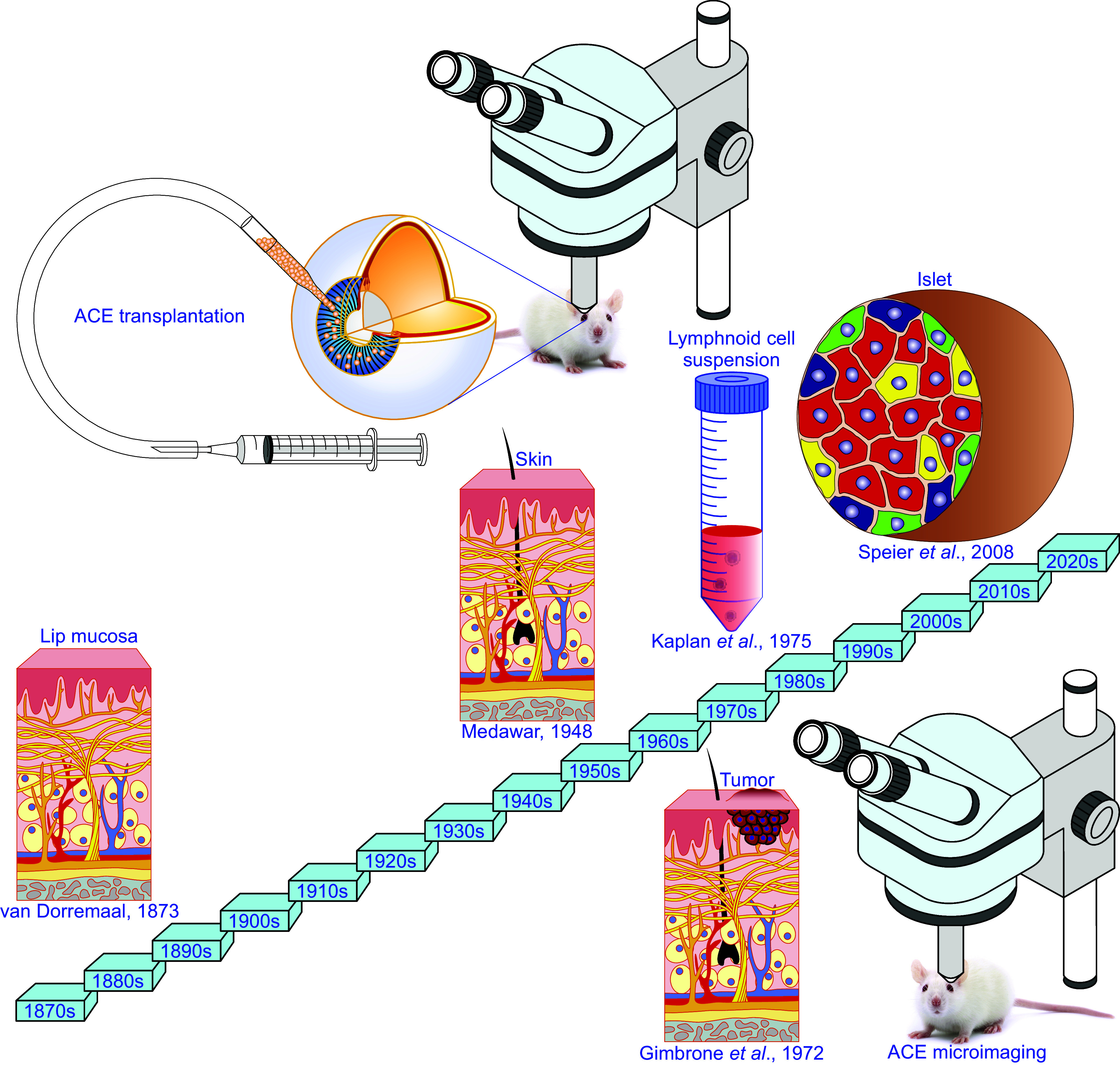
    1. van Dooremaal JC. Die entwickelung der in fremden grund versetzten lebenden gewebe. Graefe’s Arhiv Ophthalmol 19: 359–373, 1873. doi:10.1007/BF01693910. - DOI
    1. Speier S, Nyqvist D, Cabrera O, Yu J, Molano RD, Pileggi A, Moede T, Köhler M, Wilbertz J, Leibiger B, Ricordi C, Leibiger IB, Caicedo A, Berggren PO. Noninvasive in vivo imaging of pancreatic islet cell biology. Nat Med 14: 574–578, 2008. doi:10.1038/nm1701. - DOI - PMC - PubMed
    1. Speier S, Nyqvist D, Köhler M, Caicedo A, Leibiger IB, Berggren PO. Noninvasive high-resolution in vivo imaging of cell biology in the anterior chamber of the mouse eye. Nat Protoc 3: 1278–1286, 2008. doi:10.1038/nprot.2008.118. - DOI - PMC - PubMed

LinkOut - more resources