

Percutaneous endoscopic lumbar discectomy in lumbar disc herniation with posterior ring apophysis fracture: A case report in a 15-year-old child
- PMID: 38206687
- PMCID: PMC10754556
- DOI: 10.1097/MD.0000000000036213
Percutaneous endoscopic lumbar discectomy in lumbar disc herniation with posterior ring apophysis fracture: A case report in a 15-year-old child
Abstract
Rationale: Lumbar disc herniation (LDH) with posterior ring apophysis fracture (PRAF) is rather rare in children, and in all age-stratified LDH patients, the incidence of RAF was 5.3% to 7.5%. Interestingly, the incidence of LDH with RAF in children (15%-32%) is several times higher than in adults, the mis-diagnosis of which may lead to delayed treatment.
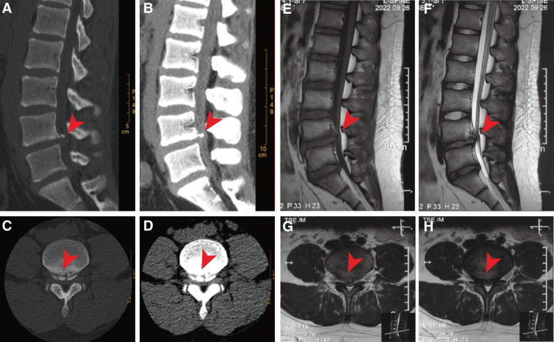
Patient concerns: Here, we report a 15-year-old schoolboy who suffered from sudden low back pain and radiating pain in both lower limbs after sport activities. Symptoms persisted after 3 months of conservative treatment. Computer radiography and magnetic resonance imaging indicated central disc herniation with PRAF at L4-5.
Diagnosis: LDH with PRAF.
Interventions: The herniated disc and epiphyseal fragments were successfully excised by the percutaneous endoscopic lumbar discectomy minimal-invasive technique.
Outcomes: Surgery was successful. Symptoms were immediately relieved postoperatively with a wound of only about 7.0 mm. Discharged on the next day. No perioperative complications occurred. Moreover, the imaging and clinical outcomes were also more satisfactory during the post-operative 15 months outpatient follow-up.
Lessons: Pediatric LDH with PRAF is extremely uncommon, and there is a lack of training among physicians for such cases, which may lead to delayed diagnosis and treatment. Once a diagnosis for LDH with PRAF is established, percutaneous endoscopic lumbar discectomy is a safe and effective minimally invasive treatment to be considered, and we hope that this technique can provide more assistance in the future.
Copyright © 2023 the Author(s). Published by Wolters Kluwer Health, Inc.
Conflict of interest statement
The authors have no funding and conflicts of interest to disclose.
Figures


Similar articles
-
Percutaneous endoscopic interlaminar discectomy for posterior ring apophyseal fracture accompanied with lumbar disc herniation in a 12-year pediatric diver: a case report.Childs Nerv Syst. 2023 Jan;39(1):275-278. doi: 10.1007/s00381-022-05605-5. Epub 2022 Jul 7. Childs Nerv Syst. 2023. PMID: 35798908 Free PMC article.
-
Percutaneous endoscopic lumbar discectomy: minimally invasive technique for multiple episodes of lumbar disc herniation.BMC Musculoskelet Disord. 2017 Aug 1;18(1):329. doi: 10.1186/s12891-017-1697-8. BMC Musculoskelet Disord. 2017. PMID: 28764746 Free PMC article.
-
[Short-term effectiveness of percutaneous endoscopic spine surgery for treatment of lumbar disc herniation with posterior ring apophysis separation].Zhongguo Xiu Fu Chong Jian Wai Ke Za Zhi. 2014 Nov;28(11):1353-7. Zhongguo Xiu Fu Chong Jian Wai Ke Za Zhi. 2014. PMID: 25639049 Chinese.
-
A review of current treatment of lumbar posterior ring apophysis fracture with lumbar disc herniation.Eur Spine J. 2013 Mar;22(3):475-88. doi: 10.1007/s00586-012-2580-9. Epub 2012 Nov 21. Eur Spine J. 2013. PMID: 23179980 Free PMC article. Review.
-
Efficacy of percutaneous endoscopic lumbar discectomy for pediatric lumbar disc herniation and degeneration on magnetic resonance imaging: case series and literature review.J Int Med Res. 2021 Jan;49(1):300060520986685. doi: 10.1177/0300060520986685. J Int Med Res. 2021. PMID: 33472475 Free PMC article. Review.
Cited by
-
Adolescent lumbar disc herniation: etiology, diagnosis, and treatment options.J Orthop Surg Res. 2025 Jun 20;20(1):605. doi: 10.1186/s13018-025-06024-3. J Orthop Surg Res. 2025. PMID: 40542380 Free PMC article. Review.
References
-
- Lavelle WF, Bianco A, Mason R, et al. . Pediatric disk herniation. J Am Acad Orthop Surg. 2011;19:649–56. - PubMed
-
- Martinez-Lage JF, Martinez Robledo A, Lopez F, et al. . Disc protrusion in the child Particular features and comparison with neoplasms. Childs Nerv Syst. 1997;13:201–7. - PubMed
-
- Yang IK, Bahk YW, Choi KH, et al. . Posterior lumbar apophyseal ring fractures: a report of 20 cases. Neuroradiology. 1994;36:453–5. - PubMed
-
- Akhaddar A, Belfquih H, Oukabli M, et al. . Posterior ring apophysis separation combined with lumbar disc herniation in adults: a 10-year experience in the surgical management of 87 cases. J Neurosurg Spine. 2011;14:475–83. - PubMed
Publication types
MeSH terms
LinkOut - more resources
Full Text Sources
Medical
Research Materials
Miscellaneous
