Clinical Trial
. 2024 May 15;229(5):1256-1265.
doi: 10.1093/infdis/jiad578.

Safety and Virologic Impact of Haploidentical NK Cells Plus Interleukin 2 or N-803 in HIV Infection

Affiliations
Clinical Trial

Safety and Virologic Impact of Haploidentical NK Cells Plus Interleukin 2 or N-803 in HIV Infection

Jeffrey S Miller et al. J Infect Dis. .

Abstract

Background: Natural killer (NK) cells are dysfunctional in chronic human immunodeficiency virus (HIV) infection as they are not able to clear virus. We hypothesized that an infusion of NK cells, supported by interleukin 2 (IL-2) or IL-15, could decrease virus-producing cells in the lymphatic tissues.

Methods: We conducted a phase 1 pilot study in 6 persons with HIV (PWH), where a single infusion of haploidentical related donor NK cells was given plus either IL-2 or N-803 (an IL-15 superagonist).

Results: The approach was well tolerated with no unexpected adverse events. We did not pretreat recipients with cyclophosphamide or fludarabine to "make immunologic space," reasoning that PWH on stable antiretroviral treatment remain T-cell depleted in lymphatic tissues. We found donor cells remained detectable in blood for up to 8 days (similar to what is seen in cancer pretreatment with lymphodepleting chemotherapy) and in the lymph nodes and rectum up to 28 days. There was a moderate decrease in the frequency of viral RNA-positive cells in lymph nodes.

Conclusions: There was a moderate decrease in HIV-producing cells in lymph nodes. Further studies are warranted to determine the impact of healthy NK cells on HIV reservoirs and if restoring NK-cell function could be part of an HIV cure strategy. Clinical Trials Registration. NCT03346499 and NCT03899480.

Keywords: HIV; IL-15; IL-2; N-803; NK cells; reservoir.


Conflict of interest statement

Potential conflicts of interest. J. T. S. and P. S. S. are affiliated with ImmunityBio, which provided N-803 for the trial. Neither of these authors had any influence on study design or data interpretation. All other authors report no potential conflicts. All authors have submitted the ICMJE Form for Disclosure of Potential Conflicts of Interest. Conflicts that the editors consider relevant to the content of the manuscript have been disclosed.

Figures

Figure 1.
Figure 1.
Schematic presentation of the clinical protocol showing the drug, timing, dose, and number of participants who received either IL-2 or N-803. There were 2 participants in the IL-2 arm, of whom 1 individual (2474) had only the postinfusion biopsy obtained. There were 4 individuals in the N-803 arm, of whom 1 individual (2611) had the baseline biopsy and we were unable to obtain the postinfusion biopsy due to COVID-19 restrictions. Abbreviations: IL-2, interleukin 2; SQ, subcutaneous.
Figure 2.
Figure 2.
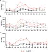
Changes in absolute numbers of (A) CD4, (B) CD8, and (C) NK cells in peripheral blood. The absolute value was determined by multiplying the absolute lymphocyte count by the percentage of lymphocytes that were either CD3+CD4+, CD3+CD8+, or CD3-CD56+ (NK cells). Abbreviations: Base, baseline; D, day; IL-2, interleukin 2; NK cell, natural killer cell; Scr, screen; W, week.
Figure 3.
Figure 3.
The frequency of CD56+ NK cells in LN, ileum, and rectum obtained one week after the last cytokine infusion. LN (A), ileum (B), and rectum (C) obtained one week after the last cytokine infusion stained with antibodies against CD56 to mark NK cells. Because there was no difference in the baseline and postinfusion frequency of CD56+ cells at each tissue site, we pooled baseline and postinfusion data to compare frequency of cells in each tissue site (D). Results from those receiving IL-2 are marked in red and those receiving N-803 in black. There was no difference between LN and ileum but the frequency in rectum was significantly lower (**P = .01, unpaired t test). Lines represent mean and standard error of the mean. Abbreviations: LN, lymph node; NK cell, natural killer cell; ns, not significant.
Figure 4.
Figure 4.
A, Adoptively transferred haploidentical NK cells persist in PBMCs up to 8 days postinfusion in 1 participant who received IL-2 (2474) and 1 who received N-803 (2611). PBMCs from each time point were stained for NK cell markers as well as a donor HLA that was missing in the recipient to look for chimerism. In the participant receiving IL-2, donor cell frequency peaked at day 6 postinfusion. In the participant receiving N-803, donor cell frequency peaked at day 4. The percentage of NK cells that are donor cells is listed in the upper right corner of the graph. Sections from lymph node (B) and rectum (image at 20x magnification) (C) from the IL-2 recipient (2474) obtained at day 21 postinfusion were stained with antibodies directed against HLA-B7, an HLA that was unique to the donor. Arrows show donor NK cells in both tissue sites, with a somewhat greater frequency in the rectum. Image at 20x magnificiation. Abbreviations: FITC, fluorescein isothiocyanate; IL-2, interleukin 2; NK cell, natural killer cell; PBMC, peripheral blood mononuclear cell; PE, phycoerythrin.
Figure 5.
Figure 5.
The frequency of (A) vRNA-positive and (B) vDNA-positive cells/g LN is shown. There was a marked and consistent reduction in the frequency of vRNA-positive cells in the 4 participants where we had paired samples but not in the frequency of vDNA-positive cells/g. Abbreviations: D, day; LN, lymph node; vDNA, viral DNA; vRNA, virial RNA.

Comment in

References

    1. Finzi D, Blankson J, Siliciano JD, et al. Latent infection of CD4+ T cells provides a mechanism for lifelong persistence of HIV-1, even in patients on effective combination therapy. Nat Med 1999; 5:512–7. - PubMed
    1. Maldarelli F, Wu X, Su L, et al. HIV latency. Specific HIV integration sites are linked to clonal expansion and persistence of infected cells. Science 2014; 345:179–83. - PMC - PubMed
    1. Wagner TA, McLaughlin S, Garg K, et al. HIV latency. Proliferation of cells with HIV integrated into cancer genes contributes to persistent infection. Science 2014; 345:570–3. - PMC - PubMed
    1. Estes JD, Kityo C, Ssali F, et al. Defining total-body AIDS-virus burden with implications for curative strategies. Nat Med 2017; 23:1271–6. - PMC - PubMed
    1. Fletcher CV, Staskus K, Wietgrefe SW, et al. Persistent HIV-1 replication is associated with lower antiretroviral drug concentrations in lymphatic tissues. Proc Natl Acad Sci U S A 2014; 111:2307–12. - PMC - PubMed

Publication types

Associated data