. 2024 May 2;143(18):1825-1836.
doi: 10.1182/blood.2023019706.

Sonrotoclax overcomes BCL2 G101V mutation-induced venetoclax resistance in preclinical models of hematologic malignancy

Affiliations

Sonrotoclax overcomes BCL2 G101V mutation-induced venetoclax resistance in preclinical models of hematologic malignancy

Jiuyang Liu et al. Blood. .

Abstract

Venetoclax, the first-generation inhibitor of the apoptosis regulator B-cell lymphoma 2 (BCL2), disrupts the interaction between BCL2 and proapoptotic proteins, promoting the apoptosis in malignant cells. Venetoclax is the mainstay of therapy for relapsed chronic lymphocytic leukemia and is under investigation in multiple clinical trials for the treatment of various cancers. Although venetoclax treatment can result in high rates of durable remission, relapse has been widely observed, indicating the emergence of drug resistance. The G101V mutation in BCL2 is frequently observed in patients who relapsed treated with venetoclax and sufficient to confer resistance to venetoclax by interfering with compound binding. Therefore, the development of next-generation BCL2 inhibitors to overcome drug resistance is urgently needed. In this study, we discovered that sonrotoclax, a potent and selective BCL2 inhibitor, demonstrates stronger cytotoxic activity in various hematologic cancer cells and more profound tumor growth inhibition in multiple hematologic tumor models than venetoclax. Notably, sonrotoclax effectively inhibits venetoclax-resistant BCL2 variants, such as G101V. The crystal structures of wild-type BCL2/BCL2 G101V in complex with sonrotoclax revealed that sonrotoclax adopts a novel binding mode within the P2 pocket of BCL2 and could explain why sonrotoclax maintains stronger potency than venetoclax against the G101V mutant. In summary, sonrotoclax emerges as a potential second-generation BCL2 inhibitor for the treatment of hematologic malignancies with the potential to overcome BCL2 mutation-induced venetoclax resistance. Sonrotoclax is currently under investigation in multiple clinical trials.


Conflict of interest statement

Conflict-of-interest disclosure: All authors are current or previous employees and shareholders of BeiGene (Beijing) Co, Ltd. The design of, conduct of, and financial support for this research were provided by BeiGene (Beijing) Co, Ltd. BeiGene (Beijing) Co, Ltd, participated in data interpretation and the review and approval of the manuscript.

Figures

None
Graphical abstract
Figure 1.
Figure 1.
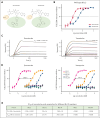
Sonrotoclax is a second-generation BCL2 inhibitor with superior potency and selectivity. (A) Chemical structures of sonrotoclax and venetoclax. The differences are highlighted with light yellow and green ellipses. (B) Cell-free competitive binding assays were performed to measure the disruption of the interaction between the BCL2:BAK-derived peptide by sonrotoclax and venetoclax. (C) SPR binding curves of sonrotoclax and venetoclax. The lines in different colors are the actual curves for the serial concentrations of sonrotoclax (0.625-10 nM) and venetoclax (3.125-200 nM), whereas the corresponding black lines are the fitted curves using the 1:1 binding model. KD values are presented as the mean values ± standard deviations (SDs) for 4 independent experiments. (D) Inhibition of BCL-xL, BCL-W, MCL-1, and BCL2A1 was measured with a method similar to that described in (B). (E) Measured IC50 values of sonrotoclax and venetoclax for the inhibition of Bcl-2 family members. The data are presented as the mean values ± SDs of 3 independent experiments.
Figure 2.
Figure 2.
Sonrotoclax is more efficacious than venetoclax in both cancer cells and xenograft mouse models. (A) Cell viability inhibition was measured using a CellTiter-Glo luminescence assay. (B) Disruption of the BCL2:BIM complex was detected by a cell-based competitive Meso Scale Discovery (MSD) assay. (C) The activation of caspase-3 and caspase-7 (caspase-3/7) was measured in RS4;11 cells with a Caspase-Glo kit. Annexin V+ cells were quantified by FITC annexin V staining and FACS analysis in RS4;11 cells. The accumulation of sub-G0/G1 in RS4;11 cells was assessed by PI staining and FACS analysis. The data are presented as the mean values ± SDs for 3 independent experiments, with representative plots shown in the figures. EC50, half maximal effective concentration. (D) MV4-11 (AML), MAVER-1 (MCL), and Toledo (DLBCL) cells were treated with serial dilutions of sonrotoclax or venetoclax for 2 days, and cell viability inhibition was measured by a CellTiter-Glo luminescence assay. IC50 values are presented as the mean values ± SDs for 3 independent experiments. (E-G) The in vivo antitumor activity of sonrotoclax and venetoclax was evaluated in human RS4;11 (E), MAVER-1 (F), and Toledo (G) xenograft models. Mice were treated with sonrotoclax or venetoclax once daily at the indicated doses by oral gavage. The tumor volumes are presented as the mean values ± standard error of the mean (SEMs) of 8 (E) or 10 (F-G) mice in each group; ∗∗∗∗P < .0001. EC50, half maximal effective concentration, FACS, fluorescence-activated cell sorting; FITC, fluorescein isothiocyanate; mpk, milligrams per kilogram; PI, propidium iodide.
Figure 3.
Figure 3.
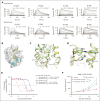
Molecular mechanism of the interaction between BCL2 and sonrotoclax. (A) Overview of the structure of BCL2 in complex with sonrotoclax. The protein is shown as an electrostatic surface, with the compound shown in yellow. (B) Binding details of sonrotoclax. Water molecules are shown as red spheres. Hydrogen bonds are represented by green dashed lines. (C) Superimposition of the structures of BCL2 in complex with sonrotoclax (yellow stick) and venetoclax (cyan stick, protein shown as pink ribbon, PDB ID: 6O0K). (D) Zoomed in view of the superimposed structures shown in panel C. The F112 residues underneath the P2 pocket are shown as purple and pink sticks. (E) Comparison of the occupation of the P2 pocket by venetoclax (cyan stick) and sonrotoclax. The subpocket is indicated by a red arrow. (F) Zoomed in view of the binding details of the (S)-2-(2-isopropylphenyl)pyrrolidine moiety. The subpocket is encircled by a light orange dashed line, and the sulfonyl-π and π-π interactions are indicated by purple dashed lines.
Figure 4.
Figure 4.
Sonrotoclax is effective against the venetoclax-resistant BCL2 G101V mutant. (A) Binding affinities of sonrotoclax (left panel) and venetoclax (right panel) to the BCL2 G101V mutant measured by SPR. The curves in different colors represent the serial concentrations of sonrotoclax (0.625-20 nM) and venetoclax (6.25-800 nM). KD values are presented as the mean values ± SDs for 4 independent experiments. (B) The disruption of the cellular BCL2:BIM complex in BCL2 G101V KI RS4;11 cells by sonrotoclax and venetoclax were detected using the MSD assay. (C) Inhibition of the cell viability in the parental RS4;11 and G101V KI cells was evaluated by a CellTiter-Glo luminescence assay. IC50 values are presented as the mean values ± SDs for 3 independent experiments, with representative curves shown in panels B and C. (D-E) Assessment of in vivo PD/PK and efficacy of sonrotoclax and venetoclax in RS4;11 G101V xenograft models. Mice were treated with sonrotoclax or venetoclax at the indicated doses. Single-dose treatment was used to evaluate PD, and daily dosing was used for the efficacy study; all treatments were administered by oral gavage. For evaluation of PD/PK, plasma and tumor tissues were collected at the specified time points to measure drug concentrations and tumor cleaved caspase-3 (Asp175) levels by ELISA. For the efficacy study, the tumor volume was measured twice weekly, and the TGI was calculated on day 18 after dosing. The data are presented as the mean values ± SEMs of 3 mice at each time point (D) and 8 mice in each group (E); ∗∗∗∗P < .0001. ELISA, enzyme linked immunosorbent assay; PD, pharmacodynamics; PK, pharmacokinetics.
Figure 5.
Figure 5.
Crystal structure of the BCL2 G101V mutant in complex with sonrotoclax. (A) Comparison of the structures of WT BCL2 (yellow) and the G101V mutant (gray) in complex with sonrotoclax. (B-C) Conformational changes of residues around G/V101 in the crystal structures of WT BCL2 and G101V in complex with venetoclax (B) and sonrotoclax (C). The complex structures of BCL2:venetoclax (PDB ID: 6O0K) and BCL2 G101V:venetoclax (PDB ID: 6O0L) are shown in cyan and light orange, respectively. The minimum distances between E152 of the BCL2 G101V mutant and the compounds are indicated by green and red dashed lines.
Figure 6.
Figure 6.
Sonrotoclax remains effective against other BCL2 mutants. (A) SPR measurements of the binding affinities of sonrotoclax and venetoclax to different BCL2 variants. The curves in different colors represent different concentrations of sonrotoclax (0.156-20 nM for D103Y, 0.313-20 nM for V156D, 0.078-20 nM for A113G, and 1.25-20 nM for R129L) and venetoclax (0.781-200 nM for D103Y, 1.56-100 nM for V156D, 0.781-400 nM for A113G, and 0.781-200 nM for R129L). KD values are presented as the mean values ± SDs for 4 independent experiments. (B) Mutations are indicated on the BCL2 protein surface (gray). The mutated residues are highlighted in cyan. Sonrotoclax is represented by the yellow stick. (C) Superimposition of the structures of WT BCL2 (yellow) and the D103Y mutant (pale green) in complex with sonrotoclax. (D) Zoomed in view of D103 or Y103 involved in the binding of sonrotoclax or venetoclax. The hydrogen bonds are indicated by the cyan and yellow dashed lines. (E) Inhibition of the viability of parental and BCL2 D103Y KI KMS-12-PE cells was estimated by the CellTiter-Glo luminescence assay. IC50 values are presented as the mean values ± SDs for 3 independent experiments, with representative curves shown in the figure. (F) Efficacy evaluation of sonrotoclax and venetoclax in the KMS-12-PE D103Y xenograft model. The tumor volumes are presented as the mean values ± SEMs of 8 animals in each group; ∗∗P < .01.
Figure 7.
Figure 7.
Schematic diagram of the knockon effect of the V101 side chain against E152. (A) G101V mutation interferes with BCL2-venetoclax interaction through E152. (B) G101V mutation is too distant from sonrotoclax to impede its binding, even via E152. The legends are shown below the diagrams.

Comment in

  • BCL2 inhibition: back to the future!
    Dyer MJS, Walter HS. Dyer MJS, et al. Blood. 2024 May 2;143(18):1787-1788. doi: 10.1182/blood.2023023796. Blood. 2024. PMID: 38696193 No abstract available.

References

    1. Fernald K, Kurokawa M. Evading apoptosis in cancer. Trends Cell Biol. 2013;23(12):620–633. - PMC - PubMed
    1. Elmore S. Apoptosis: a review of programmed cell death. Toxicol Pathol. 2007;35(4):495–516. - PMC - PubMed
    1. Tsujimoto Y, Finger LR, Yunis J, Nowell PC, Croce CM. Cloning of the chromosome breakpoint of neoplastic B cells with the t(14;18) chromosome translocation. Science. 1984;226(4678):1097–1099. - PubMed
    1. Hockenbery D, Nuñez G, Milliman C, Schreiber RD, Korsmeyer SJ. Bcl-2 is an inner mitochondrial membrane protein that blocks programmed cell death. Nature. 1990;348(6299):334–336. - PubMed
    1. Hockenbery DM, Zutter M, Hickey W, Nahm M, Korsmeyer SJ. BCL2 protein is topographically restricted in tissues characterized by apoptotic cell death. Proc Natl Acad Sci U S A. 1991;88(16):6961–6965. - PMC - PubMed

Publication types

MeSH terms