Review
. 2024 Apr;29(4):1139-1152.
doi: 10.1038/s41380-023-02399-z. Epub 2024 Jan 12.

Iron imbalance in neurodegeneration

Affiliations
Review

Iron imbalance in neurodegeneration

Sonia Levi et al. Mol Psychiatry. 2024 Apr.

Abstract

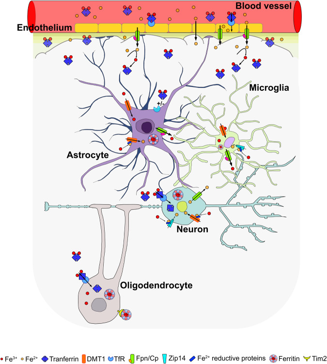
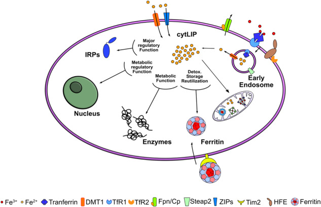
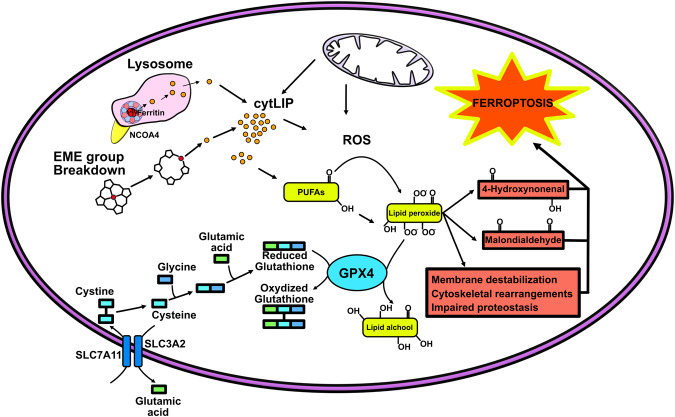
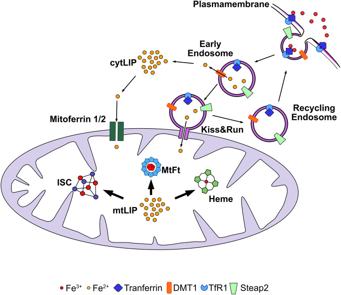
Iron is an essential element for the development and functionality of the brain, and anomalies in its distribution and concentration in brain tissue have been found to be associated with the most frequent neurodegenerative diseases. When magnetic resonance techniques allowed iron quantification in vivo, it was confirmed that the alteration of brain iron homeostasis is a common feature of many neurodegenerative diseases. However, whether iron is the main actor in the neurodegenerative process, or its alteration is a consequence of the degenerative process is still an open question. Because the different iron-related pathogenic mechanisms are specific for distinctive diseases, identifying the molecular mechanisms common to the various pathologies could represent a way to clarify this complex topic. Indeed, both iron overload and iron deficiency have profound consequences on cellular functioning, and both contribute to neuronal death processes in different manners, such as promoting oxidative damage, a loss of membrane integrity, a loss of proteostasis, and mitochondrial dysfunction. In this review, with the attempt to elucidate the consequences of iron dyshomeostasis for brain health, we summarize the main pathological molecular mechanisms that couple iron and neuronal death.


Conflict of interest statement

The authors declare no competing interests.

Figures

Fig. 1
Fig. 1. Cartoon depicting an example of iron transfer among different resident CNS cells and the different transporters involved.
Iron enters BBB endothelial cells as Tf-TfR1 or via NTBI binding-mediated endocytosis. The ferric ion is thus released at the basolateral side by Fpn in the CNS interstitial fluid and associates with Tf, synthesized in the choroid plexus. NTBI is associated with ascorbate, citrate or ATP (released by astrocytes). Astrocytes internalize iron via DMT1, store it in ferritin, and distribute it to cells in the CNS via Ceruloplasmin-coupled Ferroportin (Cp/Fpn). Oligodendrocytes acquire metal through the ferritin receptor Tim-2 or DMT1. Neurons can acquire iron through the Tf-TfR1 pathway and DMT1.
Fig. 2
Fig. 2. Main metabolic cellular pathways involved in iron homeostasis, usage, and transport.
Iron incorporated into the cell, via Tf/TfR1 endocytosis or through DMT1/ZIPs, reaches the cytosol and mitochondria for support the ISC and heme biosyntheses. TfR2 form a complex with hemochromatosis protein, HFE, and serves as a component of the iron sensing machinery to regulate iron homeostasis. Fpn is the only iron-protein exporter involved in release of metal from the cell. The cytosolic labile iron pool (cytLIP), the redox-active iron available for the synthesis of iron enzymes, is in direct contact with only two classes of cytosolic proteins. They are highly represented and can bind iron: ferritins bind Fe-oxygen complexes, while IRPs link Fe-S (ISC) complexes. Ferritins store excess iron, and IRPs act as iron sensors.
Fig. 3
Fig. 3. Graphic representation of the cellular mechanisms involved in the increase in ferroptotic events.
Ferroptosis leads to membrane destabilization, mitochondrial dysfunction, cytoskeletal rearrangements, and protein impairment. It is triggered by an imbalance between lipid hydroperoxide detoxification and iron-dependent ROS accumulation. The peroxidation of Polyunsaturated fatty acids (PUFAs) is limited by glutathione peroxidase 4 (GPX4), which utilizing glutathione (GSH), converts the lipid hydroperoxide in lipid alcohol. When equilibrium is lost, the oxidized lipid species (4-Hydroxynonenal and Malondialdehyde) accumulate in membranes, destabilizing them and leading to cell death. SLC7A11, solute carrier family 7 member 11 and SLC3A2, solute carrier family 3 member 2 allow the internalization of cystine need for GSH synthesis. A key ferroptotic player is glutathione depletion and/or the inactivation of glutathione-dependent antioxidant enzyme GPX4. Source of iron are heme and cytosolic ferritin degradation. Under conditions of iron restriction, NCOA4 binds to the H-subunit of ferritin, carrying it to lysosomes (ferritinophagy), where the protein is degraded and iron is released; during iron excess, NCOA4 is degraded by the ubiquitin–proteasome system, making cytosolic ferritin free to sequester iron.
Fig. 4
Fig. 4. Cartoon depicting an example of iron uptake and utilization in mitochondria.
Clathrin-coated endosomes containing TfR1-bound iron are endocytosed. The endosome lumen is acidified by a proton pump; the acidification decreases Tf-iron binding affinity, and as consequence, iron is released into the endosome lumen. Here, ferric ions are reduced by Steap2 and released through DMT1 into the cell cytosol. TfR1 is recycled back to the plasma membrane by recycling endosomes. Cytosolic free iron enters mitochondria through the mitoferrin channels. A second mechanism, called Kiss&Run, has been described to deliver iron to mitochondria, which consists of transient fusion between endosomes and mitochondrial membranes. Inside the mitochondrion, the labile iron pool (mitLIP), the redox-active form of iron, is used for sustaining heme and ISC biosynthesis or stored in mitochondrial ferritin (mtFt).

References

    1. Rouault TA. Iron metabolism in the CNS: Implications for neurodegenerative diseases. Nat Rev Neurosci. 2013;14:551–64. doi: 10.1038/nrn3453. - DOI - PubMed
    1. Gutteridge JMC, Halliwell B. Mini-Review: Oxidative stress, redox stress or redox success? Biochem Biophys Res Commun. 2018;502:183–6. doi: 10.1016/j.bbrc.2018.05.045. - DOI - PubMed
    1. Galaris D, Barbouti A, Pantopoulos K. Iron homeostasis and oxidative stress: An intimate relationship. Biochim Biophys Acta - Mol Cell Res. 2019;1866:118535. doi: 10.1016/j.bbamcr.2019.118535. - DOI - PubMed
    1. Muckenthaler MU, Galy B, Hentze MW. Systemic iron homeostasis and the iron-responsive element/iron-regulatory protein (IRE/IRP) regulatory network. Annu Rev Nutr. 2008;28:197–213. doi: 10.1146/annurev.nutr.28.061807.155521. - DOI - PubMed
    1. Hentze MW, Muckenthaler MU, Galy B, Camaschella C. Two to tango: regulation of mammalian iron metabolism. Cell. 2010;142:24–38. doi: 10.1016/j.cell.2010.06.028. - DOI - PubMed