. 2024 Jan 12;81(1):30.
doi: 10.1007/s00018-023-05045-7.

Knockdown of NEAT1 prevents post-stroke lipid droplet agglomeration in microglia by regulating autophagy

Affiliations

Knockdown of NEAT1 prevents post-stroke lipid droplet agglomeration in microglia by regulating autophagy

Yongli Pan et al. Cell Mol Life Sci. .

Abstract

Background: Lipid droplets (LD), lipid-storing organelles containing neutral lipids like glycerolipids and cholesterol, are increasingly accepted as hallmarks of inflammation. The nuclear paraspeckle assembly transcript 1 (NEAT1), a long non-coding RNA with over 200 nucleotides, exerts an indispensable impact on regulating both LD agglomeration and autophagy in multiple neurological disorders. However, knowledge as to how NEAT1 modulates the formation of LD and associated signaling pathways is limited.

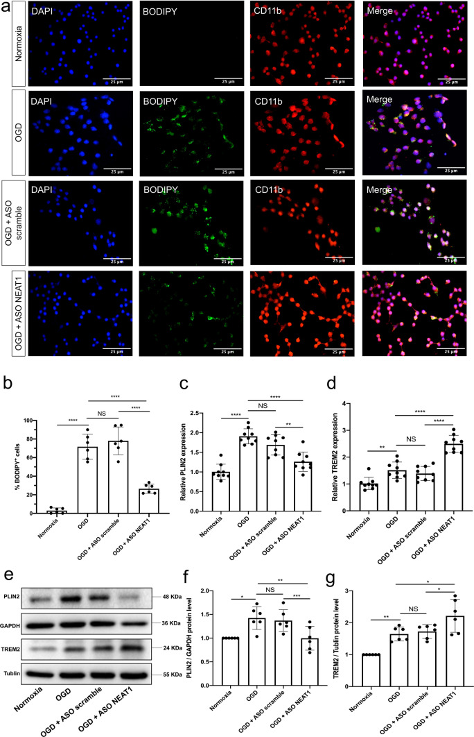
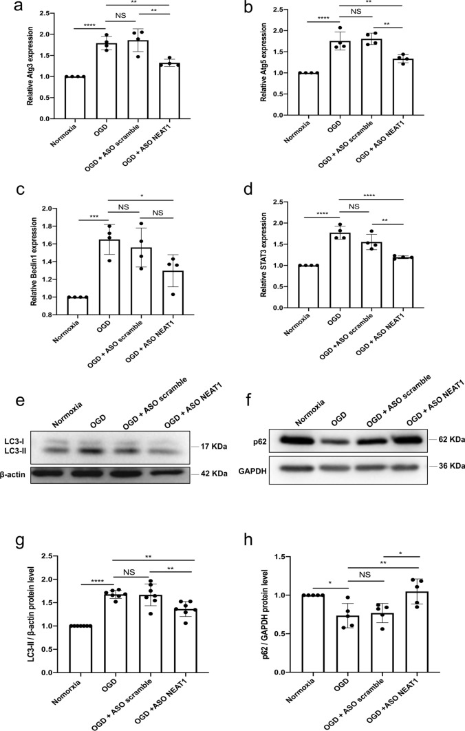
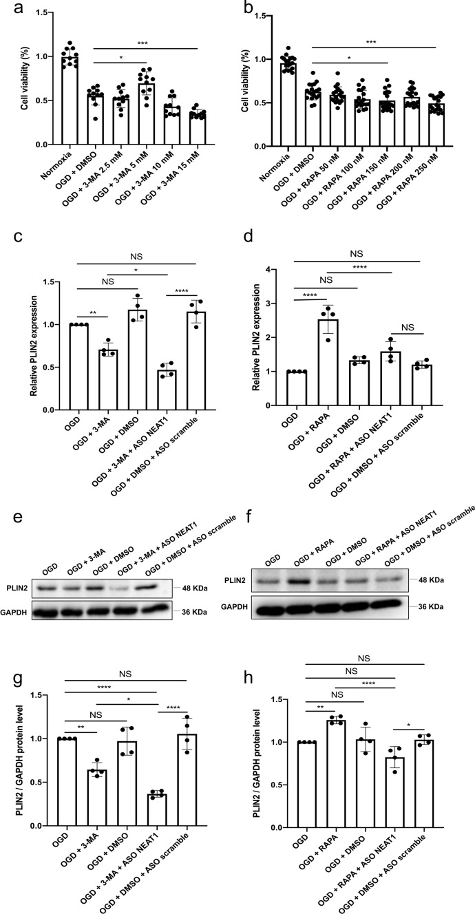
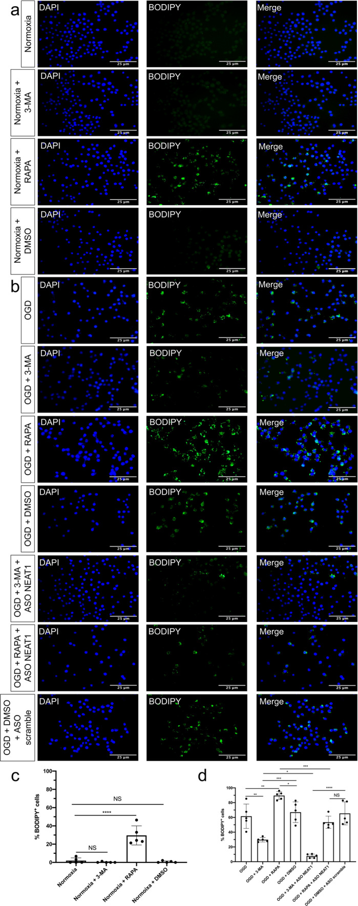
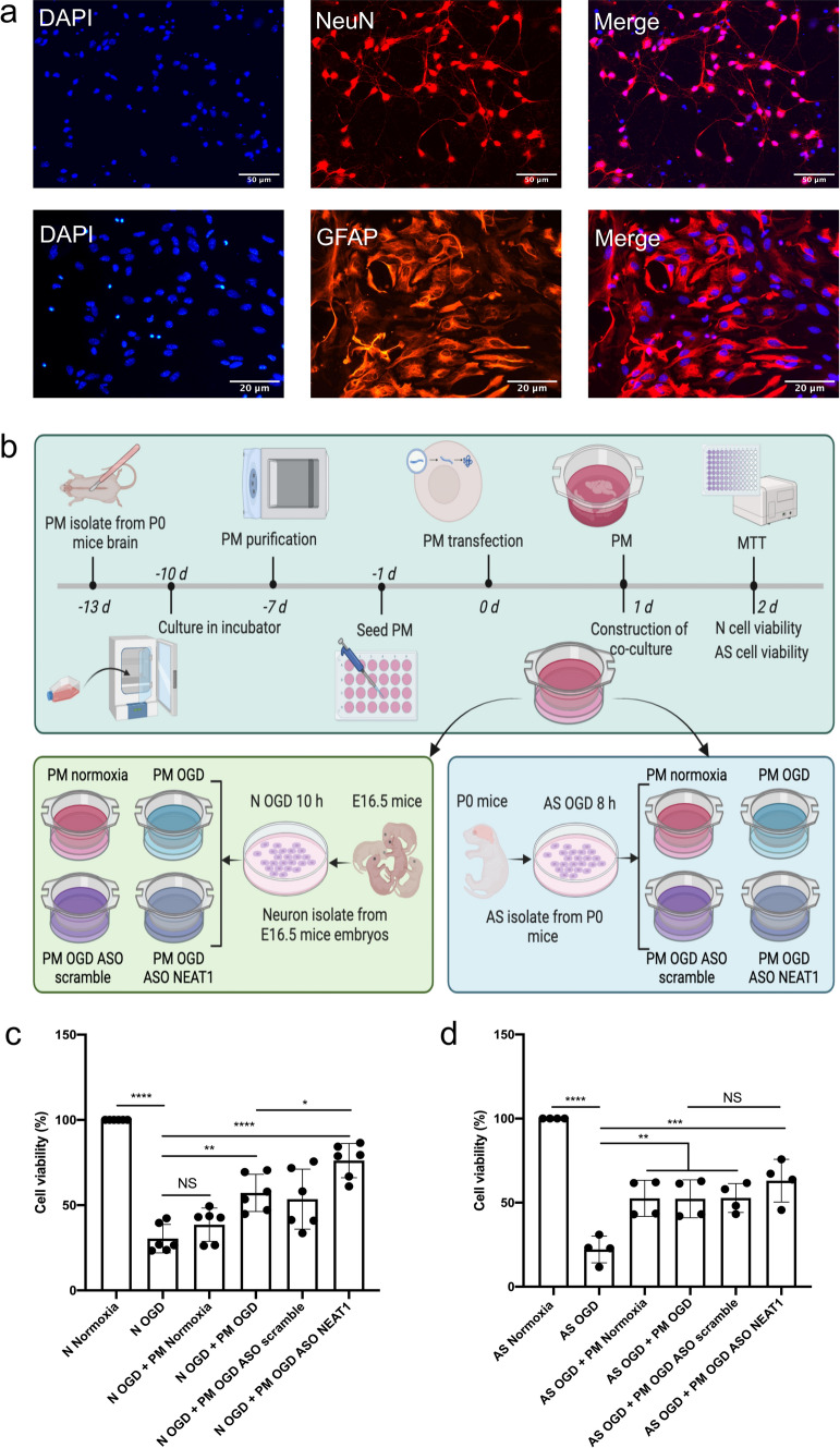
Methods: In this study, primary microglia were isolated from newborn mice and exposed to oxygen-glucose-deprivation/reoxygenation (OGD/R). To further explore NEAT1-dependent mechanisms, an antisense oligonucleotide (ASO) was adopted to silence NEAT1 under in vitro conditions. Studying NEAT1-dependent interactions with regard to autophagy and LD agglomeration under hypoxic conditions, the inhibitor and activator of autophagy 3-methyladenine (3-MA) and rapamycin (RAPA) were used, respectively. In a preclinical stroke model, mice received intraventricular injections of ASO NEAT1 or control vectors in order to yield NEAT1 knockdown. Analysis of readout parameters included qRT-PCR, immunofluorescence, western blot assays, and behavioral tests.

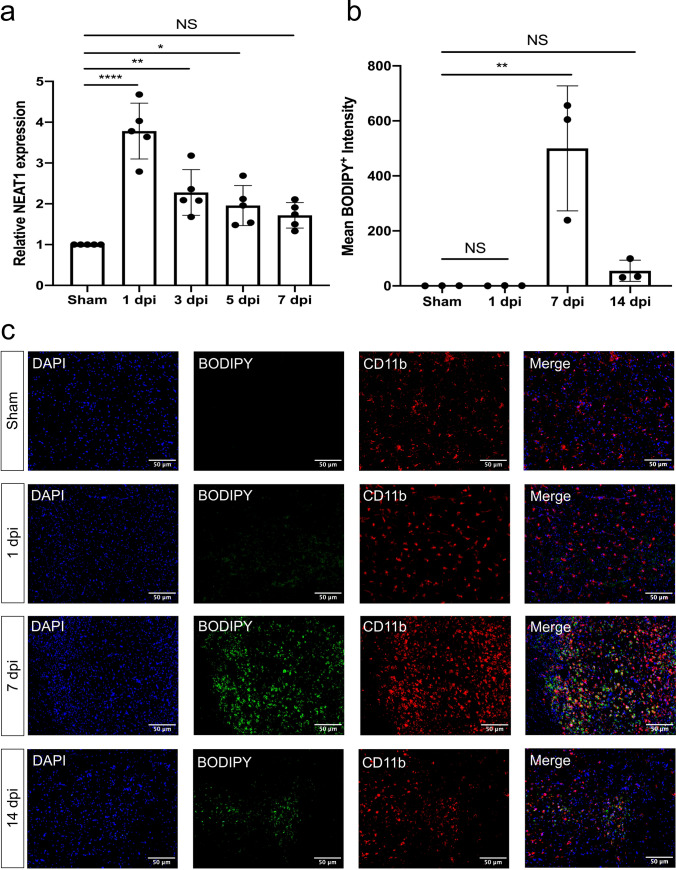
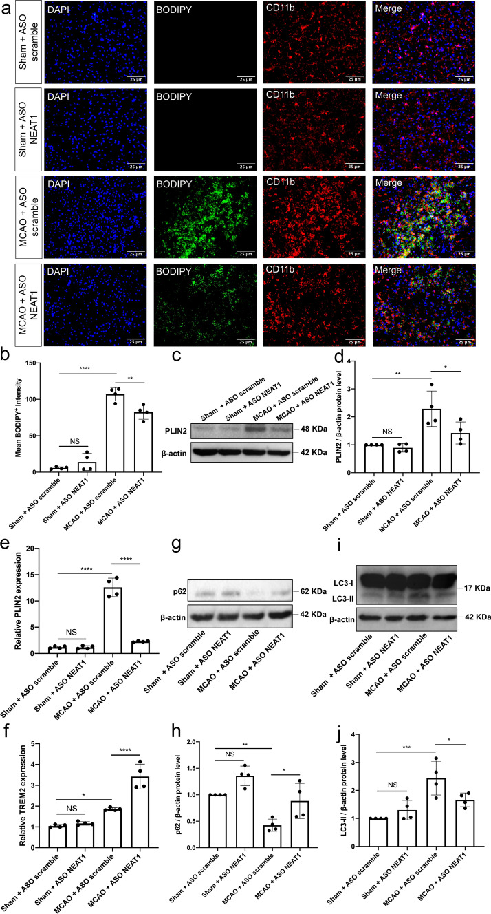
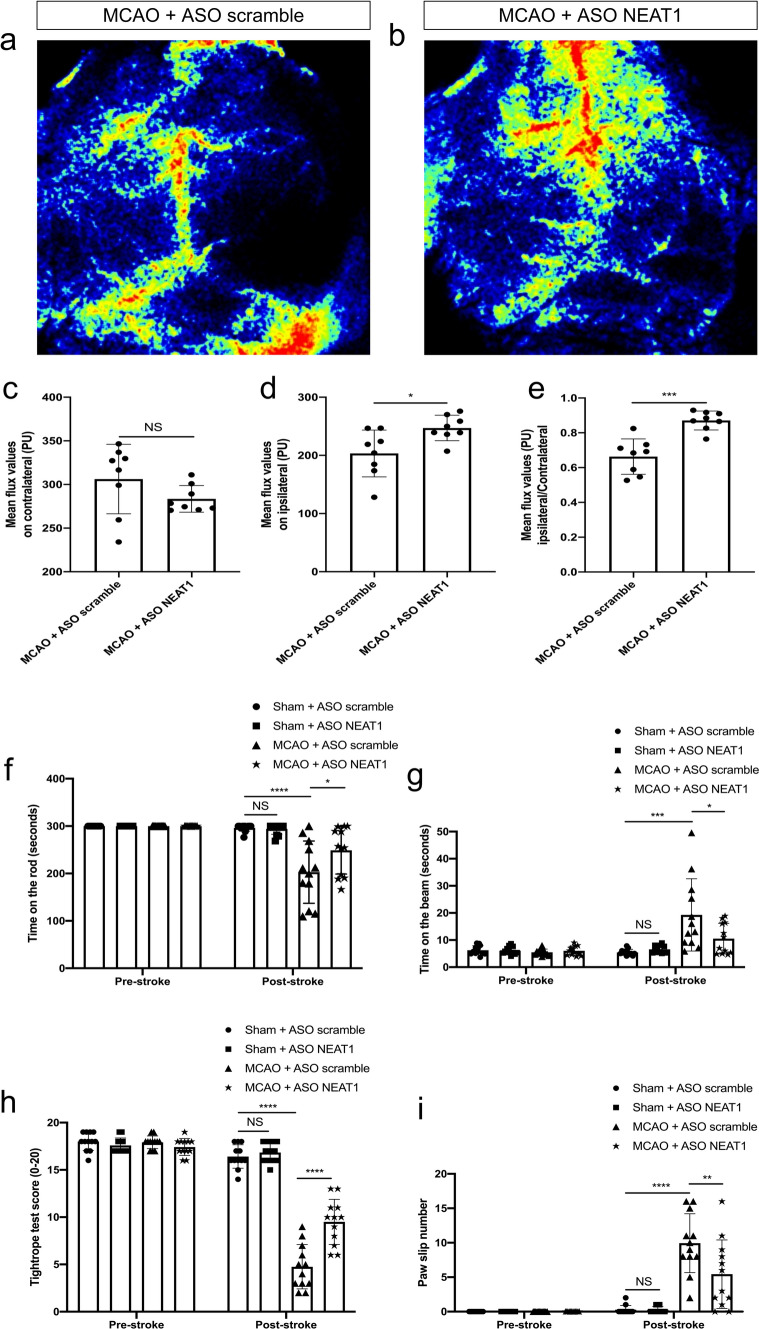
Results: Microglia exposed to OGD/R displayed a temporal pattern of NEAT1 expression, peaking at four hours of hypoxia followed by six hours of reoxygenation. After effectively silencing NEAT1, LD formation and autophagy-related proteins were significantly repressed in hypoxic microglia. Stimulating autophagy in ASO NEAT1 microglia under OGD/R conditions by means of RAPA reversed the downregulation of LD agglomeration and perilipin 2 (PLIN2) expression. On the contrary, application of 3-MA promoted repression of both LD agglomeration and expression of the LD-associated protein PLIN2. Under in vivo conditions, NEAT1 was significantly increased in mice at 24 h post-stroke. Knockdown of NEAT1 significantly alleviated LD agglomeration and inhibited autophagy, resulting in improved cerebral perfusion, reduced brain injury and increased neurological recovery.

Conclusion: NEAT1 is a key player of LD agglomeration and autophagy stimulation, and NEAT1 knockdown provides a promising therapeutic value against stroke.

Keywords: Autophagy; Lipid droplets; Microglia; NEAT1; Stroke.


Conflict of interest statement

None of the authors has a financial relationship with a commercial entity that has an interest in the subject of this manuscript.

Figures

Fig. 1
Fig. 1
Light microscopy of primary microglia and exposure towards OGD with subsequent NEAT1 expression and cell viability analyses. Primary microglia were isolated from cerebral cortices and hippocampi of newborn WT C57BL/6 J mice. a Light microscopy of primary microglia in culture. b The magnified image of primary microglia under bright-field microscopy. c, d The corresponding NEAT1 expression at different time points of OGD and reoxygenation in comparison to PPIA. e, f The MTT assay was used to evaluate the cell viability after various OGD and reoxygenation time points. All results are expressed as mean ± standard deviation and analyzed by one-way ANOVA followed by Tukey's post-hoc-test. NS, no significance, **p < 0.01, ***p < 0.001, ****p < 0.0001. Abbreviations: NEAT1, nuclear paraspeckle assembly transcript 1; OGD, oxygen-glucose-deprivation; PPIA, peptidylprolyl isomerase A
Fig. 2
Fig. 2
Knockdown of NEAT1 affects expression patterns of LD in primary microglia exposed to hypoxia. a The fluorescence staining of neutral LD (BODIPY, green) in microglial cells (CD11b, red) cultured with ASO scramble or ASO NEAT1. b Statistical analysis of the percentage of LD in whole cells. c, d Primary microglia were transfected with ASO NEAT1 (or ASO scramble as control) for 24 h to generate a stable NEAT1 knockdown. The expression of the LD-related genes PLIN2 (c) and TREM2 (d) was detected using qRT-PCR in comparison to PPIA. eg Western blot analysis of PLIN2 and TREM2 expression in OGD/R microglial cells after treatment with ASO scramble or ASO NEAT1. The relative quantitative analysis was normalized against GAPDH or tublin, as indicated. All results are expressed as mean ± standard deviation and analyzed by one-way ANOVA followed by Tukey's post-hoc-test. NS, no significance, *p < 0.05, **p < 0.01, ***p < 0.001, and ****p < 0.0001. Abbreviations: NEAT1, nuclear paraspeckle assembly transcript 1; ASO, antisense oligonucleotide; OGD/R, oxygen-glucose-deprivation/reoxygenation; LD, lipid droplets; PLIN2, perilipin 2; TREM2, triggering receptor expressed on myeloid cells 2; PPIA, peptidylprolyl isomerase A
Fig. 3
Fig. 3
Knockdown of NEAT1 affects signaling cascades related to autophagy in primary microglia exposed to hypoxia. ad Autophagy-related genes, i.e., Atg3 (a), Atg5 (b), Beclin1 (c), and STAT3 (d) were determined by quantitative qRT-PCR normalized to PPIA. eh NEAT1 knockdown repressed LC3 but activated p62 expressions. Western blot was utilized to analyze the expression of LC3 (e) at a 17 kDa band and p62 (f) at a 62 kDa band in microglial cells in four parallel groups (normoxia, OGD, OGD + ASO scramble, and OGD + ASO NEAT1). The relative quantitative analysis was normalized against β-actin or GAPDH. Results are expressed as mean ± standard deviation and analyzed by one-way ANOVA followed by Tukey's post-hoc-test. NS, no significance, *p < 0.05, **p < 0.01, ***p < 0.001, and ****p < 0.0001. Abbreviations: NEAT1, nuclear paraspeckle assembly transcript 1; ASO, antisense oligonucleotide; OGD, oxygen-glucose-deprivation; Atg3, autophagy-related 3; PPIA, peptidylprolyl isomerase A
Fig. 4
Fig. 4
Regulation of autophagy affects PLIN2 expression in primary microglia exposed to OGD. a, b The impact of autophagy on microglia survival after OGD was evaluated using different concentrations of the autophagy inhibitor 3‐MA or autophagy stimulator RAPA in comparison to microglia treated with the solvent DMSO using the MTT assay. c, d Relative PLIN2 expressions in microglial cells are shown after inhibition (c) or activation (d) of autophagy under OGD conditions using qRT-PCR normalized to PPIA. eh Protein levels of PLIN2 in microglia are displayed after inhibition or activation of the autophagy pathway under OGD conditions. PLIN2 protein was recognized as a 48 kDa band, and GAPDH (36 kDa) was used as a reference. All results are expressed as mean ± standard deviation and analyzed by one-way ANOVA followed by Tukey's post-hoc-test. NS, no significance, *p < 0.05, **p < 0.01, ***p < 0.001, and ****p < 0.0001. Abbreviations: ASO, antisense oligonucleotide; OGD, oxygen-glucose-deprivation; 3-MA, 3-methyladenine; RAPA, rapamycin; PLIN2, perilipin 2; DMSO, dimethyl sulfoxide; PPIA, peptidylprolyl isomerase A
Fig. 5
Fig. 5
Regulation of autophagy affects LD formation in primary microglia. a Microglial cells were treated with 3-MA or RAPA under normoxia conditions, followed by BODIPY staining. b Microglia were treated with 3-MA or RAPA with additional ASO scramble or ASO NEAT1 treatment under hypoxia conditions, followed by BODIPY staining. c, d Quantification analysis of the percentage of LD in whole cells. All results are expressed as mean ± standard deviation and analyzed by one-way ANOVA followed by Tukey's post-hoc-test. NS, no significance, *p < 0.05, **p < 0.01, ***p < 0.001, and ****p < 0.0001. Abbreviations: LD, lipid droplets; 3-MA, 3-methyladenine; RAPA, rapamycin; ASO, antisense oligonucleotide; NEAT1, nuclear paraspeckle assembly transcript 1
Fig. 6
Fig. 6
Knockdown of NEAT1 in microglia protects primary neurons from OGD injury. a Immunofluorescence images of primary neurons (NeuN) and astrocytes (GFAP) in red under normoxia conditions. Nuclei were counterstained with DAPI in blue. b A summary of the experimental paradigm for the in vitro co-culture model. Primary microglia and astrocytes were isolated from neonatal C57BL/6 mice on postnatal day 0, whereas primary cortical neurons were obtained from E16.5 mouse embryos. The neurons were subjected to 10 h of OGD, whereas astrocytes underwent 8 h of OGD. Microglia were subjected to four conditions (normoxia, OGD, OGD + ASO scramble, OGD + ASO NEAT1), followed by co-culture with hypoxic neuorns or astrocytes. Afterwards, they were cultured under normoxic conditions for 24 h. c, d The MTT assay was performed to assess the cell survival of primary neurons and astrocytes co-cultured with microglia, which were treated in different ways. As such, microglia were treated with ASO NEAT1 knockdown constructs or with scramble constructs. All results are expressed as mean ± standard deviation and analyzed by one-way ANOVA followed by Tukey's post-hoc-test. NS, no significance, *p < 0.05, **p < 0.01, ***p < 0.001, and ****p < 0.0001. Abbreviations: NEAT1, nuclear paraspeckle assembly transcript 1; ASO, antisense oligonucleotide; OGD, oxygen-glucose-deprivation; PM, primary microglia; N, neuron; AS, astrocyte
Fig. 7
Fig. 7
NEAT1 and LD expression in stroke mice. a The expression of NEAT1 on different days following cerebral ischemia in comparison to PPIA. b Quantification of the fluorescence intensity expression of BODIPY. c Images show the dual staining for BODIPY and CD11b in the ischemic hemisphere taken from sham and different days post-ischemic reperfusion. Results are expressed as mean ± standard deviation and analyzed by one-way ANOVA followed by Tukey's post-hoc-test. NS, no significance, *p < 0.05, **p < 0.01, and ****p < 0.0001. Abbreviations: NEAT1, nuclear paraspeckle assembly transcript 1; dpi, day post-ischemia; PPIA, peptidylprolyl isomerase A
Fig. 8
Fig. 8
NEAT1 knockdown represses LD agglomeration and inhibits autophagy following MCAO in vivo. Mice were exposed to 45 min of MCAO followed by 7 days of survival. Sham group mice underwent the surgery procedure without MCAO. a Images show the dual fluorescence staining for BODIPY and CD11b in the ischemic hemisphere taken from the sham group, the negative control group (MCAO + ASO scramble) or mice treated with ASO NEAT1 at 7 dpi. Quantification of the mean BODIPY+ fluorescence intensity expressions between each group of mice is shown in b. c, d Western blot analysis of PLIN2 in whole ipsilateral cortex tissues and β-actin was used as a reference. ef The expression of PLIN2 and TREM2 in the ipsilateral ischemic cerebral cortex from sham-operated mice (sham + ASO scramble, sham + ASO NEAT1) or from mice at 7 dpi (MCAO + ASO scramble, MCAO + ASO NEAT1) was examined by qRT-PCR. gj Representative images of western blots are shown for the expression of p62 (g) and LC3 (i) in whole ipsilateral cortex tissues. Bar graphs show the quantitative analyses of western blots as ratios of p62/β-actin (h) and LC3/β-actin (j). All results are expressed as mean ± standard deviation and analyzed by one-way ANOVA (n = 4 mice per experimental group) followed by Tukey's post-hoc-test. NS, no significance, *p < 0.05, **p < 0.01, ***p < 0.001, and ****p < 0.0001. Abbreviations: LD, lipid droplets; ASO, antisense oligonucleotide; MCAO, middle cerebral artery occlusion; PLIN2, perilipin 2; TREM2, triggering receptor expressed on myeloid cells 2; NEAT1, nuclear paraspeckle assembly transcript 1; dpi, day post-ischemia
Fig. 9
Fig. 9
Silencing NEAT1 protects against ischemia-induced brain damage. All mice were exposed to 45 min of MCAO. a, b Laser speckle images show the effect of ASO scramble or ASO NEAT1 on cortical blood circulation at 7dpi. ce Statistical analysis of laser speckle measurements is shown for the contralateral cortex, the ipsilateral cortex, and for the ratio ipsilateral to contralateral in mice exposed to MCAO at 7dpi. fi Knockdown of NEAT1 reduces post-ischemic motor coordination impairment, which was evaluated using the test of rotarod (f), the balance beam (g), the tightrope (h), and the paw slip (i) on day 1 before the stroke and day 7 after the stroke. All results are expressed as mean ± standard deviation and analyzed by Student's t-test or two-way ANOVA (n = 8 mice per experimental group for laser speckle measurements and 12 mice for behavior tests) followed by Tukey's post-hoc-test. NS, no significance, *p < 0.05, **p < 0.01, ***p < 0.001, and ****p < 0.0001. Abbreviations: ASO, antisense oligonucleotide; MCAO, middle cerebral artery occlusion; NEAT1, nuclear paraspeckle assembly transcript 1; dpi, day post-ischemia

References

    1. Woodburn S, Bollinger J, Wohleb E. The semantics of microglia activation: neuroinflammation, homeostasis, and stress. J Neuroinflammation. 2021;18(1):258. doi: 10.1186/s12974-021-02309-6. - DOI - PMC - PubMed
    1. He T, et al. Sestrin2 regulates microglia polarization through mTOR-mediated autophagic flux to attenuate inflammation during experimental brain ischemia. J Neuroinflammation. 2020;17(1):329. doi: 10.1186/s12974-020-01987-y. - DOI - PMC - PubMed
    1. Lu X, et al. Novel therapeutic strategies for ischemic stroke: recent insights into autophagy. Oxid Med Cell Longev. 2022;2022:3450207. doi: 10.1155/2022/3450207. - DOI - PMC - PubMed
    1. Hu K, et al. Review of the effects and mechanisms of microglial autophagy in ischemic stroke. Int Immunopharmacol. 2022;108:108761. doi: 10.1016/j.intimp.2022.108761. - DOI - PubMed
    1. Alarcon-Gil J, et al. Neuroprotective and anti-inflammatory effects of linoleic acid in models of parkinson's disease: the implication of lipid droplets and lipophagy. Cells. 2022;11(15):1. doi: 10.3390/cells11152297. - DOI - PMC - PubMed