. 2024 Jan 11;22(1):46.
doi: 10.1186/s12967-023-04819-8.

ANGPTL4 accelerates ovarian serous cystadenocarcinoma carcinogenesis and angiogenesis in the tumor microenvironment by activating the JAK2/STAT3 pathway and interacting with ESM1

Affiliations

ANGPTL4 accelerates ovarian serous cystadenocarcinoma carcinogenesis and angiogenesis in the tumor microenvironment by activating the JAK2/STAT3 pathway and interacting with ESM1

Yu-Kun Li et al. J Transl Med. .

Abstract

Background: Ovarian cancer (OC) is a malignant neoplasm that displays increased vascularization. Angiopoietin-like 4 (ANGPTL4) is a secreted glycoprotein that functions as a regulator of cell metabolism and angiogenesis and plays a critical role in tumorigenesis. However, the precise role of ANGPTL4 in the OC microenvironment, particularly its involvement in angiogenesis, has not been fully elucidated.

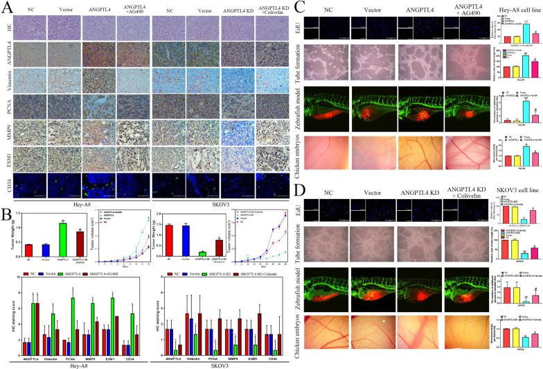
Methods: The expression of ANGPTL4 was confirmed by bioinformatics and IHC in OC. The potential molecular mechanism of ANGPTL4 was measured by RNA-sequence. We used a series of molecular biological experiments to measure the ANGPTL4-JAK2-STAT3 and ANGPTL4-ESM1 axis in OC progression, including MTT, EdU, wound healing, transwell, xenograft model, oil red O staining, chick chorioallantoic membrane assay and zebrafish model. Moreover, the molecular mechanisms were confirmed by Western blot, Co-IP and molecular docking.

Results: Our study demonstrates a significant upregulation of ANGPTL4 in OC specimens and its strong association with unfavorable prognosis. RNA-seq analysis affirms that ANGPTL4 facilitates OC development by driving JAK2-STAT3 signaling pathway activation. The interaction between ANGPTL4 and ESM1 promotes ANGPTL4 binding to lipoprotein lipase (LPL), thereby resulting in reprogrammed lipid metabolism and the promotion of OC cell proliferation, migration, and invasion. In the OC microenvironment, ESM1 may interfere with the binding of ANGPTL4 to integrin and vascular-endothelial cadherin (VE-Cad), which leads to stabilization of vascular integrity and ultimately promotes angiogenesis.

Conclusion: Our findings underscore that ANGPTL4 promotes OC development via JAK signaling and induces angiogenesis in the tumor microenvironment through its interaction with ESM1.

Keywords: ANGPTL4; Angiogenesis; ESM1; JAK2/STAT3 pathway; Ovarian cancer.


Conflict of interest statement

The authors declare no competing interests.

Figures

Fig. 1
Fig. 1
The expression, potential functions, diagnostic value and prognostic value of ARGs in OC patients. A The mRNA expression of ARGs in OC samples compared to normal ovary samples based on the TCGA database. B The diagnostic value of ARGs for OC patients. C The PPI network for ARGs. D The correlation among these ARGs in OC patients. E GO and F KEGG enrichment analysis for these ARGs. G ANGPTL4 protein expression in OC and normal ovaries based on the HPA database. H The prognostic value of ANGPTL4 for OC patients based on the TCGA database. I The expression of ANGPTL4 in normal ovary samples and benign and malignant ovarian disease samples. J GSEA of ANGPTL4 based on the TCGA database. K Immune infiltration analysis for OC patients according to ANGPTL4 expression. L The expression of ANGPTL4 in multiple OC cell lines. M The expression of ESM1 and ANGPTL4 in multiple OC cell lines by Western blot. *P < 0.05, **P < 0.01, ***P < 0.001
Fig. 2
Fig. 2
The effects of the ANGPTL4/JAK2/STAT3 pathway on OC proliferation, migration and invasion. A The protein levels of ANGPTL4, p-JAK2, JAK2, p-STAT3, STAT3, Vimentin, PCNA and hypoxia-inducible factor alpha (HIF-α) were detected in the NC-SKOV3, Vector-SKOV3, ANGPTL4 KD-SKOV3, ANGPTL4 KD-SKOV3 with Colivelin, NC-HeyA8, Vector-HeyA8, ANGPTL4 OE-HeyA8, and ANGPTL4 OE-HeyA8 with AG490 groups by Western blotting. B A workflow for transfecting cells, collecting conditioned medium, and conducting analyses on OC cells and HUVECs. The effect of ANGPTL4, ANGPTL4 + AG490, ANGPTL4 KD, and ANGPTL4 KD + Colivelin on Hey-A8 and SKOV3 cell proliferation ability via MTT (C) and EdU (D) analysis. The migration and invasion abilities of ANGPTL4, ANGPTL4 + AG490, ANGPTL4 KD, and ANGPTL4 KD + Colivelin were measured by wound healing (E) and Transwell invasion assays (F) in Hey-A8 and SKOV3 cells, respectively. *P < 0.05, **P < 0.01, ***P < 0.001
Fig. 3
Fig. 3
ANGPTL4 promotes OC growth and angiogenesis by activating the JAK2/STAT3 pathway in vivo. A Morphological observation and HE and IHC staining of xenografts in the NC-SKOV3, Vector-SKOV3, ANGPTL4 KD-SKOV3, ANGPTL4 KD-SKOV3 with Colivelin, NC-HeyA8, Vector-HeyA8, ANGPTL4 OE-HeyA8, and ANGPTL4 OE-HeyA8 with AG490 groups. B The tumor weight and volume and the IHC staining score of xenografts in the NC-SKOV3, Vector-SKOV3, ANGPTL4 KD-SKOV3, ANGPTL4 KD-SKOV3 with Colivelin, NC-HeyA8, Vector-HeyA8, ANGPTL4 OE-HeyA8, and ANGPTL4 OE-HeyA8 with AG490 groups. C The proliferation and tube formation of HUVECs cultured with the CM of HeyA8 cells were tested by EdU staining and tube formation assays. The chick chorioallantoic membrane assay measured the angiogenesis ability of HeyA8 CM in vivo. Angiogenesis ability of HeyA8 cells in a zebrafish model. The experimental groups were NC, Vector, ANGPTL4 OE and ANGPTL4 + AG490. D The proliferation and tube formation of HUVECs cultured with the CM of SKOV3 cells were tested by EdU staining and tube formation assays. The chick chorioallantoic membrane assay measured the angiogenesis ability of SKOV3 CM in vivo. Angiogenesis ability of SKOV3 cells in the zebrafish model. The experimental groups were NC, Vector, ANGPTL4 KD and ANGPTL4 KD + Colivelin. *P < 0.05, **P < 0.01, ***P < 0.001
Fig. 4
Fig. 4
ANGPTL4 interacted with ESM1. A The correlation expression of ESM1 and ANGPTL4 in OC samples based on TCGA database. B IF staining for ANGPTL4 and ESM1 in OC tissue samples, SKOV3 cell lines, HeyA8 cell lines and HUVECs. C The interaction of ANGPTL4 and ESM1 in HeyA8 cells by Co-IP analysis. D GST pull-down analysis for GST-ANGPTL4 and His-ESM1. E Molecular docking for ANGPTL4 and ESM1 by HADDOCK.
Fig. 5
Fig. 5
ESM1 promotes ANGPTL4 to combine with LPL by accelerating proliferation, invasion and lipid accumulation in OC cells. A Molecular docking for ANGPTL4, LPL and ESM1 by HADDOCK. B Co-IP showed the effects of ESM1 on the interaction between ANGPTL4 and LPL in HeyA8 and SKOV3 cells. The effect of vector, ANGPTL4, ANGPTL4 + ESM1, ANGPTL4 KD, and ANGPTL4 KD + ESM1 on Hey-A8 and SKOV3 cell lipid levels and proliferation ability via oil red O staining (C), MTT (D) and EdU (E) analysis. The migration and invasion abilities were measured by wound healing (F) and Transwell assays (G) in Hey-A8 and SKOV3 cells transfected with vector, ANGPTL4, ANGPTL4 + ESM1, ANGPTL4 KD, and ANGPTL4 KD + ESM1. *P < 0.05, **P < 0.01, ***P < 0.001
Fig. 6
Fig. 6
ESM1 inhibits ANGPTL4 binding to integrin α5β1 and VE-cadherin, which represses HUVEC proliferation and migration to induce vascular permeability in the tumor microenvironment. A Co-IP showed that ESM1 inhibited ANGPTL4 binding to α5β1 and VE-cadherin in HUVECs cultured with HeyA8 and SKOV3 cells, respectively. The effect of vector, ANGPTL4, ANGPTL4 + ESM1, ANGPTL4 KD, and ANGPTL4 KD + ESM1 on HUVECs cultured with Hey-A8 and SKOV3 cell lipid levels and proliferation ability via oil red O staining (B), MTT (C) and EdU (D) analysis. The migration ability was measured by wound healing (F) and Transwell assays (G) in HUVECs cultured with Hey-A8 and SKOV3 cells transfected with vector, ANGPTL4, ANGPTL4 + ESM1, ANGPTL4 KD, and ANGPTL4 KD + ESM1. *P < 0.05, **P < 0.01, ***P < 0.001
Fig. 7
Fig. 7
A model of the molecular mechanism by which ANGPTL4 promotes OC development and progression. A OC is a type of tumor with a rich blood supply. B There are many angiogenic factors in the tumor microenvironment of OC. C A variety of cells in the tumor microenvironment can take up these angiogenesis factors in a variety of ways to produce different pathophysiological effects. D ANGPTL4 can promote the proliferation and migration of OC cells by activating the JAK-STAT3 pathway, and ESM1 can bind ANGPTL4 and stabilize the binding of ANGPTL4 to LPL, thus promoting lipid accumulation and accelerating the proliferation and migration of OC cells. ANGPTL4 can also promote endothelial cell proliferation and migration through the JAK-STAT3 signaling pathway. ESM1 can inhibit the binding of ANGPTL4 to integrin α5β1 to increase vascular stability. Moreover, ESM1 promotes angiogenesis in the ovarian cancer microenvironment by binding to ANGPTL4 to inhibit its destruction of VE-cadherin-mediated tight junctions. E The molecular mechanism might be attributed to the interaction of ESM1 and ANGPTL4.

References

    1. Gómez-Raposo C, Mendiola M, Barriuso J, Casado E, Hardisson D, Redondo A. Angiogenesis and ovarian cancer. Clin Transl Oncol. 2009;11:564–571. doi: 10.1007/s12094-009-0406-y. - DOI - PubMed
    1. Shaw D, Clamp A, Jayson GC. Angiogenesis as a target for the treatment of Ovarian cancer. Curr Opin Oncol. 2013;25:558–565. doi: 10.1097/CCO.0b013e328363e0da. - DOI - PubMed
    1. Perren TJ, Swart AM, Pfisterer J, Ledermann JA, Pujade-Lauraine E, Kristensen G, Carey MS, Beale P, Cervantes A, Kurzeder C, et al. A phase 3 trial of bevacizumab in ovarian cancer. N Engl J Med. 2011;365:2484–2496. doi: 10.1056/NEJMoa1103799. - DOI - PubMed
    1. Kong DH, Kim MR, Jang JH, Na HJ, Lee S. A review of anti-angiogenic targets for monoclonal antibody cancer therapy. Int J Mol Sci. 2017;18:1786. doi: 10.3390/ijms18081786. - DOI - PMC - PubMed
    1. Tan MJ, Teo Z, Sng MK, Zhu P, Tan NS. Emerging roles of angiopoietin-like 4 in human cancer. Mol Cancer Res. 2012;10:677–688. doi: 10.1158/1541-7786.MCR-11-0519. - DOI - PubMed

Publication types

LinkOut - more resources