. 2024 Jan 11;21(1):18.
doi: 10.1186/s12974-024-03010-0.

Lipoxins A4 and B4 inhibit glial cell activation via CXCR3 signaling in acute retinal neuroinflammation

Affiliations

Lipoxins A4 and B4 inhibit glial cell activation via CXCR3 signaling in acute retinal neuroinflammation

Izhar Livne-Bar et al. J Neuroinflammation. .

Abstract

Lipoxins are small lipids that are potent endogenous mediators of systemic inflammation resolution in a variety of diseases. We previously reported that Lipoxins A4 and B4 (LXA4 and LXB4) have protective activities against neurodegenerative injury. Yet, lipoxin activities and downstream signaling in neuroinflammatory processes are not well understood. Here, we utilized a model of posterior uveitis induced by lipopolysaccharide endotoxin (LPS), which results in rapid retinal neuroinflammation primarily characterized by activation of resident macroglia (astrocytes and Müller glia), and microglia. Using this model, we observed that each lipoxin reduces acute inner retinal inflammation by affecting endogenous glial responses in a cascading sequence beginning with astrocytes and then microglia, depending on the timing of exposure; prophylactic or therapeutic. Subsequent analyses of retinal cytokines and chemokines revealed inhibition of both CXCL9 (MIG) and CXCL10 (IP10) by each lipoxin, compared to controls, following LPS injection. CXCL9 and CXCL10 are common ligands for the CXCR3 chemokine receptor, which is prominently expressed in inner retinal astrocytes and ganglion cells. We found that CXCR3 inhibition reduces LPS-induced neuroinflammation, while CXCR3 agonism alone induces astrocyte reactivity. Together, these data uncover a novel lipoxin-CXCR3 pathway to promote distinct anti-inflammatory and proresolution cascades in endogenous retinal glia.

Keywords: CXCL10; CXCL9; CXCR3; Gliosis; Inflammation resolution; Lipoxins; Neuroinflammation; Retina; Uveitis.


Conflict of interest statement

Several authors (I. L-B, J.M.S., K.G.) have obtained a US patent for use of lipoxins to treat neurodegeneration.

Figures
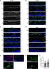
Fig. 1
Fig. 1
Intravitreal LPS induces rapid activation of inner retinal glial cells. LPS was injected intravitreally and several markers of resident and infiltrating inflammatory cells were monitored over 8 days. A Increased Müller cell and astrocyte reactivity in the inner retina was detected with the marker GFAP in glial fibers (arrows) by day 2. B Staining with the macrophage marker F4-80 (green) indicated a marked increase of infiltrating cells at the vitreo-retinal interface (arrows) in the inner retina by day 2, as well as activated microglia (asterisks). C Iba1 staining for microglial density was generally consistent within the retina across sections (asterisks), but also highlighted the appearance of vitreo-retinal macrophages (arrows). D GR-1 positive neutrophils infiltrated into the vitreous by 24 h after LPS (d1) where they accumulated at the inner retinal surface (green; arrows). However, GR-1 positive cells were never substantially observed within the retina, and had largely disappeared by 2 days post LPS injection (d2–d8). E Representative higher magnification images of the markers used (scale bars represent 20 µm). F Representative retinal Iba-1 stained images of ramified or amoeboid microglial morphology in the LPS-treated group (scale bar represents 20 µm). G Quantification of total inner-retina Iba1-positive cells on day 2 shows no significant change in macrophage/microglia numbers in the LPS group compared to vehicle (bars represent SE). (GCL; ganglion cell layer, INL; inner nuclear layer, scale bars of AD represent 100 µm)
Fig. 2
Fig. 2
Pretreatment with LXA4 or LXB4 reduces astrocyte and Müller cell reactivity in the inner retina. A Schematic of intravitreal pretreatment and subsequent LPS retinal inflammation assessment at 48 h. B Representative images showing pretreatment with LXA4 or LXB4 strongly reduced GFAP-positive Müller glia fibers in the inner retina (GFAP, arrows). C Pretreatment with LXA4 or LXB4 did not appear to reduce LPS-induced activated microglia (amoeboid Iba1; green, arrows) compared to vehicle control (Veh). D Treatment with LXA4 or LXB4 did not appear to alter the levels of infiltrating macrophages (F4-80: green) at the vitreo-retinal interface (arrows). E Corresponding quantification of activated Müller glia in the inner retina expressed as the number of GFAP + processes. LXA4 and LXB4 pretreatment significantly reduced LPS-activated Müller glia compared with vehicle (Veh, *p < 0.05, **p < 0.01). F Quantification of activated (amoeboid) microglia from the inner retina showed no significant reduction with LXA4 or LXB4 pretreatment compared to vehicle (Veh). G Infiltrating macrophages were quantified at the vitreo-retinal interface, showing no significant effect of LXA4 and LXB4 treatment. (GCL; ganglion cell layer, IPL; inner plexiform layer, INL; inner nuclear layer, scale bars represent 100 µm, graph bars represent SE)
Fig. 3
Fig. 3
Therapeutic treatment with LXB4 reduces microglial activation in the inner retina. A Schematic of the LPS-induced retinal inflammation model, subsequent intravitreal treatments and evaluation at 72 h (48 h after lipoxin treatments). B Representative images of therapeutic treatment with LXA4 or LXB4 suggesting little effect on Müller glia activation highlighted by GFAP staining of retinal fibers (arrows). C Activated microglia (amoeboid Iba1; green, arrows) in the inner retina were reduced by post-inflammation treatment with LXA4 or LXB4. D Therapeutic treatment with LXA4 or LXB4 did not substantially reduce the levels of infiltrating macrophages (F4-80; green) accumulating at the vitreal–retinal interface (arrows). E Corresponding quantification of activated Müller glia confirmed that LXA4 and LXB4 post-inflammation treatment had no significant effect compared to Vehicle (Veh). F Quantification of activated amoeboid microglia in the inner retina showed that post-inflammation LXB4 treatment significantly reduced activated microglia compared to control (**p < 0.01 G) Quantification of F4-80 staining confirmed no significant effect of LXA4 or LXB4 treatment post-inflammation. (GCL; ganglion cell layer, IPL; inner plexiform layer, INL; inner nuclear layer, scale bars represent 100 µm, graph bars represent SE)
Fig. 4
Fig. 4
CXCL9/CXCL10 induction is inhibited by LXA4 and LXB4 treatment and may interact with the CXCR3 receptor in the inner retina. A Heatmap of mean multiplex array results for 32 cytokines showing log2 fold inhibition of LPS-induced levels by LXA4 or LXB4. CXCL9 and CXCL10 were the top hits inhibited by each lipoxin (NC; not calculated due to levels being out of range). B Concentrations of CXCL9 and CXCL10 were significantly increased in retinal extracts at 24 h following LPS challenge compared to vehicle. However, eyes pre-treated with LXA4 or LXB4 showed significantly reduced induction of CXCL9 or CXCL10 levels compared to vehicle (*p < 0.05, **p < 0.01, ***p < 0.005). C The shared CXCL9/CXCL10 receptor, CXCR3 (green) is localized to the inner retina (arrows), and appeared induced at two days following LPS challenge. D CXCR3 staining partially co-localizes with the RGC marker Brn3a (red, arrows), and E CXCR3 (green) also partially co-localizes with GFAP (red, arrows). (Scale bars represent 100 µm, graph bars represent SE)
Fig. 5
Fig. 5
CXCR3 signaling modulates resident retinal inflammation. A Representative images indicating treatment with the CXCR3 antagonist AMG487 reduced Müller fiber staining at 48 h following LPS-challenge (green; arrows). Corresponding quantification showed significantly reduced Müller glia activation (*p < 0.05). B Treatment with AMG487 reduced amoeboid microglia activation (Iba1; green, arrows). Corresponding quantification of amoeboid microglia showed that AMG487 treatment strongly reduced microglia activation compared to vehicle (***p < 0.0005). C AMG487 treatment reduced LPS-induced macrophage infiltration (F4-80; green, arrows), but corresponding quantification showed a trend that did not reach significance. D To confirm the microglial results staining was also performed for CD68, which showed a similarly significantly reduced presence of amoeboid cells following AMG487 treatment. E In contrast, treatment with the CXCR3 agonist VUF11222 strongly induced astrocyte and Müller glia reactivity (GFAP, arrows). Corresponding quantification showed a significant increase in relative GFAP intensity in the inner retina in the agonist treated group compared to vehicle. F VUF11222 treatment had no effect on microglial activation. G A cartoon outlining a proposed signaling pathway by which lipoxins inhibit neuroinflammatory responses. (GCL; ganglion cell layer, IPL; inner plexiform layer, INL; inner nuclear layer, bars represent 100 µm, graph bars represent SE)

Similar articles

Cited by

References

    1. Kwon HS, Koh SH. Neuroinflammation in neurodegenerative disorders: the roles of microglia and astrocytes. Transl Neurodegener. 2020;9(1):42. doi: 10.1186/s40035-020-00221-2. - DOI - PMC - PubMed
    1. Wyss-Coray T, Mucke L. Inflammation in neurodegenerative disease—a double-edged sword. Neuron. 2002;35(3):419–432. doi: 10.1016/S0896-6273(02)00794-8. - DOI - PubMed
    1. Alqawlaq S, Flanagan JG, Sivak JM. All roads lead to glaucoma: induced retinal injury cascades contribute to a common neurodegenerative outcome. Exp Eye Res. 2019;183:88–97. doi: 10.1016/j.exer.2018.11.005. - DOI - PubMed
    1. Sivak JM. The aging eye: common degenerative mechanisms between the Alzheimer's brain and retinal disease. Invest Ophthalmol Vis Sci. 2013;54(1):871–880. doi: 10.1167/iovs.12-10827. - DOI - PubMed
    1. Liu B, Teschemacher AG, Kasparov S. Astroglia as a cellular target for neuroprotection and treatment of neuro-psychiatric disorders. Glia. 2017;65(8):1205–1226. doi: 10.1002/glia.23136. - DOI - PMC - PubMed