. 2022 Aug 18;11(4):225-236.
doi: 10.4103/jmau.jmau_62_21. eCollection 2023 Oct-Dec.

Comparative Histological Study of Therapeutic Effect of Mesenchymal Stem Cells versus Mesenchymal Stem Cells Co-Cultured with Liver Tissue on Carbon Tetrachloride-Induced Hepatotoxicity in Adult Male Albino Rats

Affiliations

Comparative Histological Study of Therapeutic Effect of Mesenchymal Stem Cells versus Mesenchymal Stem Cells Co-Cultured with Liver Tissue on Carbon Tetrachloride-Induced Hepatotoxicity in Adult Male Albino Rats

Bothina Abo-Alazm Salem et al. J Microsc Ultrastruct. .

Abstract

Context: Liver diseases are major causes of morbidity and mortality. Mesenchymal stem cells (MSCs) have immunomodulatory, anti-inflammatory, and antifibrotic effects, so they can be used in the treatment of liver diseases. MSCs co-cultured with diseased liver tissue improve the homing capacity, survival rate, and paracrine effects of the MSCs, as well as the ability to enhance liver function.

Aims: This work aimed to study the therapeutic effect of MSCs versus MSCs co-cultured with liver tissue on carbon tetrachloride (CCl4)-induced hepatotoxicity in adult male albino rats.

Settings and design: Twenty adult male albino rats were divided into four equal groups; Group I (control group), Group II received CCl4 intraperitoneally (i.p.), Group III received CCl4 i.p. and then injected with MSCs intravenously (i.v.), and Group IV received CCl4 i.p. and then injected with co-cultured MSCs i.v.

Materials and methods: Finally, liver specimens were processed for light microscopy (LM) and electron microscopy (EM). Statistical analysis was carried out to assess histological scoring, area percentage of collagen fibers, number of glial fibrillary acidic protein-positive cells, and biochemical analysis of alanine aminotransferase and aspartate aminotransferase.

Statistical analysis used: Statistical analysis of (histological scoring, area % of collagen fibers, and biochemical analysis) was done by using one-way analysis of variance (ANOVA) test using graphpad software (SanDiego, CA, USA). The means ± standard deviations were used for statistical analysis.

Results: LM of Group II revealed loss of hepatic architecture and diffuse fibrosis with dilated congested blood vessels, bile ductular proliferation, and cellular infiltrations. Vacuolated cytoplasm with or without pyknotic nuclei was observed in addition to micro- and macro-steatosis. EM demonstrated disfigured hepatocytes with abnormal organelles surrounding atypical nucleus. Group III showed restoration of the normal liver architecture with greater extent in Group IV. Statistical analysis confirmed the microscopic findings.

Conclusions: Co-cultured MSCs with diseased liver tissue augmented the therapeutic effects of MSCs in treating hepatotoxicity induced by CCl4 in adult male albino rats.

Keywords: Carbon tetrachloride; co-cultured mesenchymal stem cells; hepatotoxicity; mesenchymal stem cells.


Conflict of interest statement

There are no conflicts of interest.

Figures

Figure 1
Figure 1
Liver sections from control group showing (a) normal hepatic architecture with ill-defined classic hepatic lobules. Central vein (CV) presents in the center of the lobule and portal tracts (PT) at the periphery of the lobule (H and E, ×100). (b) Portal tract is formed of a branch of portal vein (PV), a branch of hepatic artery (bifid arrow), bile ductules (wavy arrow), lymphatics (L), and few inflammatory cells (arrowhead) (H and E, ×400). (c) Hepatocytes are arranged in anastomosing cords (thin arrows) with acidophilic cytoplasm and central rounded vesicular nuclei. Some hepatocytes are binucleated (curved arrow). Hepatocytes cords are separated by blood sinusoids (S) lined by endothelial cells with flat nuclei (angled arrows) (H and E, ×1000). Note: Kupffer cells with large, ovoid nuclei (feathery arrows)
Figure 2
Figure 2
Liver sections from hepatotoxic group showing (a) Disturbed liver architecture, with extensive deposition of thick portal and septal fibrous strands (thick arrows) (H and E, ×100). (b and c) Irregular, dilated, and congested central vein (CV), dilated portal venule (PV) and bile ductular proliferation (wavy arrows). Some hepatocytes showing vacuolated cytoplasm (V), alternating with hepatocytes with highly acidophilic cytoplasm and darkly stained nuclei (arrows) (H and E, ×400). Nucleated lightly acidophilic hepatocytes are also observed (a). Further, there is perivascular cellular infiltration (arrowhead). Note: severe necrotic area (NE). (d) Hepatocytes with many small fat droplets in their cytoplasm (micro-vesicular steatosis) (stars) (H and E, ×1000). (e) Hepatocytes showing macrovesicular steatosis (F), vacuolated cytoplasm (V), and highly acidophilic cytoplasm with dark stained nuclei (arrows) (H and E, ×1000). There is interstitial cellular infiltration (arrowheads)
Figure 3
Figure 3
Liver sections from mesenchymal stem cells-treated group (a and b) showing (a) central vein (CV) with intact endothelial lining, surrounded by cords of hepatocytes. Most of hepatocytes are normal (arrows), few cells show vacuolated cytoplasm (V) and other few cells have fragmented nuclei (H and E, ×400). (b) Portal tract with mild dilated portal venule (PV), bile ductular proliferation (wavy arrows) and few periportal inflammatory cells (arrowheads). Some hepatocytes have vacuolated cytoplasm (V) (H and E, ×400). Liver sections from co-cultured mesenchymal stem cells-treated group (c and d) showing (c) portal tract with normal bile ductules (wavy arrows) and localized perivascular cellular infiltrations (arrowhead) (H and E, ×400). (d) Normal hepatocytes (thin arrows) with acidophilic cytoplasm and vesicular nuclei (N) with prominent nuclei (H and E, ×1000). Note: Kupffer cells (feathery arrows)
Histogram 1
Histogram 1
Morphometric analysis of (a) histological scoring, (b) mean area percentage of collagen fibers, (c) number of glial fibrillary acidic protein-positive cells, (d) biochemical analysis of alanine aminotransferase, (e) biochemical analysis of aspartate aminotransferase
Figure 4
Figure 4
(a and b) Liver sections from control group showing blue-stained thin strands of collagen fibers around central vein (CV) and portal tract (PT) (Masson trichrome, ×400). (c) Liver sections from hepatotoxic group showing widespread of thick blue-stained collagen bundles at portal (double thin arrows), septal (green thick arrows), and interstitial (black thick arrows) regions giving pseudolobulation appearance (Masson trichrome, ×100). (d and e) liver section from mesenchymal stem cells-treated group showing thin blue-stained collagen fibers localized around central vein (CV) and portal tract (PT) (Masson trichrome, ×200). (f and g) Liver section from co-cultured mesenchymal stem cells-treated group showing thin blue-stained collagen fibers localized around central vein (CV) and portal tract (PT) (Masson trichrome, ×200)
Figure 5
Figure 5
Liver sections of (a) control group showing stellate moderate glial fibrillary acidic protein-positive cells along sinusoids. (b) Hepatotoxic group showing more marked numerous strong glial fibrillary acidic protein-positive cells around portal vein and along sinusoids and along fibrotic lesions. (c) Mesenchymal stem cells-treated group and (d) co-cultured mesenchymal stem cells-treated group showing results identical to control group (GFAP immunostaining, ×400)
Figure 6
Figure 6
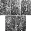
An electron micrograph of liver ultrathin sections; (a) control group showing normal binucleated hepatocyte with euchromatic nuclei (N) and prominent nucleoli (stars) surrounded by multiple mitochondria (M), dispersed rough endoplasmic reticulum (thin arrows), and glycogen granules spread in all cytoplasm (arrowheads). Note: cell junctions between hepatocytes (short arrow). (b) Hepatotoxic group showing nucleus (N) with irregularity of nuclear membrane. There is rarefaction of the cytoplasm (R) with many vacuolations (v), aggregated clusters of proliferated dilated rough endoplasmic reticulum (thin arrows), and many lysosomes (curved arrows). Note: depleted glycogen granules and prominent microvilli in bile canaliculi (wavy arrow). The inset shows proliferated dilated rough endoplasmic reticulum. (c) Hepatotoxic group showing disfigured hepatocyte having nucleus (N) with abnormal chromatin condensation and widening of peri-nuclear space (thick arrow). The cytoplasm is disorganized containing markedly dilated rough endoplasmic reticulum (thin arrow), and mitochondria with cristolysis (M). In addition, there are many vacuolations (V), lipid droplets (L), lysosomes (curved arrows), and glycogen depletion. The inset demonstrates cristolysis of the presented mitochondria. (d) Mesenchymal stem cells-treated group showing polygonal hepatocyte containing rounded euchromatic nucleus (N) with prominent nucleolus (star). The cytoplasm is well organized containing many mitochondria (m), well organized dispersed rough endoplasmic reticulum (thin arrow), lipid droplets (L), and few vacuolations (V) in association with autophagosome (A). (e) Co-cultured mesenchymal stem cells-treated group showing euchromatic binucleated hepatocyte (N) surrounded by well-organized cytoplasm. The cell also contains multiple distributed mitochondria (M), rough endoplasmic reticulum (thin arrows), and lysosomes (curved arrows) (×2500)

Similar articles

References

    1. Standring S, Anand N, Birch R, Collins P, Crossman R, Gleeson M, et al. Gray's Anatomy, The Anatomical Basis of Clinical Practice. 41st ed. Ch. 67. USA: Elsevier; 2016. Liver; pp. 1160–71.
    1. Ovalle W, Nahirney P. Netter's Essential Histology: With Correlated Histopathology. 3rd ed. Vol. 14. China: Elsevier; 2021. Liver, gallbladder, and exocrine pancreas; pp. 334–46.
    1. Pandit A, Sachdeva T, Bafna P. Drug-induced hepatotoxicity. Appl Pharm Sci. 2012;2:233–43.
    1. Margini C, Vukotic R, Brodosi L, Bernardi M, Andreone P. Bone marrow derived stem cells for the treatment of end-stage liver disease. World J Gastroenterol. 2014;20:9098–105. - PMC - PubMed
    1. Hako R, Kristian P, Jariuška P, Haková I, Hockicková I, Schréter I, et al. Noninvasive assessment of liver fibrosis in patients with chronic hepatitis B or C by contrast-enhanced magnetic resonance imaging. Can J Gastroenterol Hepatol. 2019;2019:3024630–8. - PMC - PubMed