. 2024 Jan 9;16(1):e12504.
doi: 10.1002/dad2.12504. eCollection 2024 Jan-Mar.

Polygenic risk for Alzheimer's disease is associated with neuroaxonal damage before onset of clinical symptoms

Affiliations

Polygenic risk for Alzheimer's disease is associated with neuroaxonal damage before onset of clinical symptoms

Sonja M Kagerer et al. Alzheimers Dement (Amst). .

Abstract

Introduction: Establishing valid diagnostic strategies is a precondition for successful therapeutic intervention in Alzheimer's disease (AD).

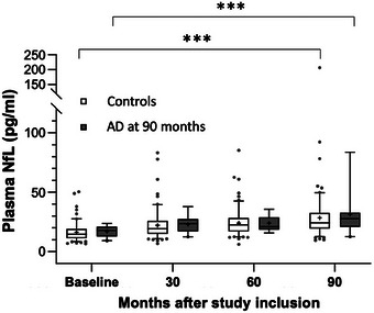
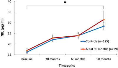
Methods: One hundred forty-four healthy 75-year-old participants from the Vienna-Transdanube-Aging longitudinal cohort study were tested for neuroaxonal damage by single molecular array (Simoa) plasma neurofilament light chain (NfL) levels at baseline, 30, 60, and 90 months, and onset of AD dementia. Individual risk for sporadic AD was estimated by continuous shrinkage polygenic risk score (PRS-CS, genome-wide association study).

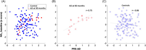
Results: Nineteen participants developed AD after a median of 60 months (interquartile range 30). In participants with AD, baseline NfL plasma levels correlated with PRS-CS (r = 0.75, p < 0.001; difference to controls: Fisher's r-to-z: z = 3.89, p < 0.001). PRS-CS combined with baseline plasma NfL predicted onset of AD (p < 0.01).

Discussion: Our data suggest that polygenic risk for AD and plasma NfL closely interact years before onset of clinical symptoms. Peripheral NfL may serve as a diagnostic measure supporting early therapeutic intervention and secondary prevention in AD.

Keywords: Alzheimer's disease; genome‐wide association studies; neurofilament light chain; plasma biomarker; polygenic risk score; single molecular array.


Conflict of interest statement

The authors declare no conflicts of interest. Author disclosures are available in the supporting information.

Figures

FIGURE 1
FIGURE 1
Plasma NfL at all time points in the AD group at 90 months and the control group. Box whisker plot of NfL (pg/mL) in plasma at 0, 30, 60, and 90 months in the AD at 90 months and the control group. Whiskers indicate 5% to 95% percentile, data points represent outliers. Differences between groups were not significant at any time point; in both groups NfL levels at 90 months were significantly different from baseline; *** p < 0.001. AD, Alzheimer's disease; NfL, neurofilament light chain.
FIGURE 2
FIGURE 2
Plasma NfL evolution over time in the AD group at 90 months and the control group. Linear plot of NfL (pg/mL; means and SEM) per time point in the AD group at 90 months and the control group. NfL levels increased significantly as a function of time (p = 0.012), independent of AD diagnosis at 90 months (p = 0.51) according to linear effects modeling. AD, Alzheimer's disease; NfL, neurofilament light chain; SEM, standard error of the mean. * p < 0.05.
FIGURE 3
FIGURE 3
Correlation of NfL at baseline with PRS‐CS in the AD at 90 months and the control group. The association of NfL at baseline (z‐scores) and PRS‐CS (z‐scores) is depicted for all participants (A), participants with AD diagnosis at 90 months (B), and the control group (C). There is a significant difference in Spearman's correlation coefficient (r) between the AD at 90 months and the control group (AD r = 0.75, controls r = −0.06, p < 0.001). Correlations are visualized by least‐squares regression line. AD, Alzheimer's disease; NfL, neurofilament light chain; PRS‐CS, polygenic risk score continuous shrinkage.

References

    1. Moscoso A, Grothe MJ, Ashton NJ, et al. Longitudinal associations of blood phosphorylated Tau181 and neurofilament light chain with neurodegeneration in Alzheimer disease. JAMA Neurol. 2021;78(4):396‐406. doi:10.1001/jamaneurol.2020.4986 - DOI - PMC - PubMed
    1. Khalil M, Teunissen CE, Otto M, et al. Neurofilaments as biomarkers in neurological disorders. Nat Rev Neurol. 2018;14(10):577‐589. doi:10.1038/s41582-018-0058-z - DOI - PubMed
    1. Kuhle J, Barro C, Andreasson U, et al. Comparison of three analytical platforms for quantification of the neurofilament light chain in blood samples: eLISA, electrochemiluminescence immunoassay and Simoa. Clin Chem Lab Med. 2016;54(10):1655‐1661. doi:10.1515/cclm-2015-1195 - DOI - PubMed
    1. Hendricks R, Baker D, Brumm J, et al. Establishment of neurofilament light chain Simoa assay in cerebrospinal fluid and blood. Bioanalysis. 2019;11(15):1405‐1418. doi:10.4155/bio-2019-0163 - DOI - PubMed
    1. Forgrave LM, Ma M, Best JR, DeMarco ML. The diagnostic performance of neurofilament light chain in CSF and blood for Alzheimer's disease, frontotemporal dementia, and amyotrophic lateral sclerosis: a systematic review and meta‐analysis. Alzheimers Dement. 2019;11:730‐743. doi:10.1016/j.dadm.2019.08.009 - DOI - PMC - PubMed