. 2024 Jan 10;16(1):e12510.
doi: 10.1002/dad2.12510. eCollection 2024 Jan-Mar.

Association of latent factors of neuroinflammation with Alzheimer's disease pathology and longitudinal cognitive decline

Affiliations

Association of latent factors of neuroinflammation with Alzheimer's disease pathology and longitudinal cognitive decline

Stefan J Teipel et al. Alzheimers Dement (Amst). .

Abstract

Introduction: We investigated the association of inflammatory mechanisms with markers of Alzheimer's disease (AD) pathology and rates of cognitive decline in the AD spectrum.

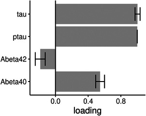
Methods: We studied 296 cases from the Deutsches Zentrum für Neurodegenerative Erkrankungen Longitudinal Cognitive Impairment and Dementia Study (DELCODE) cohort, and an extension cohort of 276 cases of the Alzheimer's Disease Neuroimaging Initiative study. Using Bayesian confirmatory factor analysis, we constructed latent factors for synaptic integrity, microglia, cerebrovascular endothelial function, cytokine/chemokine, and complement components of the inflammatory response using a set of inflammatory markers in cerebrospinal fluid.

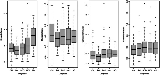
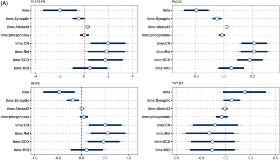
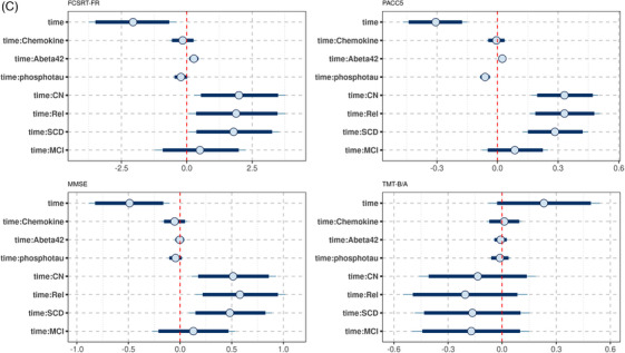
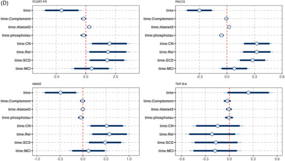
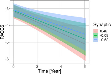
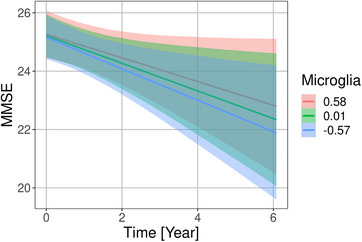
Results: We found strong evidence for an association of synaptic integrity, microglia response, and cerebrovascular endothelial function with a latent factor of AD pathology and with rates of cognitive decline. We found evidence against an association of complement and cytokine/chemokine factors with AD pathology and rates of cognitive decline.

Discussion: Latent factors provided access to directly unobservable components of the neuroinflammatory response and their association with AD pathology and cognitive decline.

Keywords: amyloid; chemokine factors; complement; endothelial function; microglia; structural equation models; tau.


Conflict of interest statement

S.J.T. participated in scientific advisory boards of Roche Pharma AG, Biogen, Grifols, and MSD, and received lecture fees from Roche and MSD. M.T.H. serves as a scientific board member of IFM Therapeutics, Novo Nordisk, and Alector and has received lecture honoraria from NovoNordisk. The other authors state no competing interests. Author disclosures are available in the supporting information.

Figures

FIGURE 1
FIGURE 1
Levels of inflammatory latent factors across groups in the DELCODE data. Boxplots of factor scores of the inflammation factors in the healthy controls (CN), the cognitively normal relatives of people with dementia (Rel), the individuals with subjective cognitive decline (SCD), and the mild cognitive impairment (MCI) and Alzheimer's disease (AD) dementia cases
FIGURE 2
FIGURE 2
Latent factor Alzheimer's disease (AD) pathology and cerebrospinal fluid (CSF) pathology markers in the DELCODE data. Association of the CSF pathology markers with the latent factor AD pathology (bars are standard deviations of the estimates) in the DELCODE data
FIGURE 3
FIGURE 3
Posterior distribution of predictors of cognitive decline in the DELCODE data. Plots of the posterior distribution of the parameter estimates for the cognitive outcomes (FCSRT‐FR, PACC5, MMSE, and TMTB/A, respectively), including effects of time and time by predictor interactions. The circle indicates the mean value, the thick segments the 90% credible intervals, and the thinner outer lines the 95% credible intervals of the posterior distribution. Of note, the time by diagnosis interactions (time:CN, controls; time:Rel, first‐degree relatives; time:SCD, subjective cognitive decline; time:MCI, mild cognitive impairment) are plotted against the reference of the AD group. So positive values for time:CN indicate that rates of cognitive decline were less pronounced in the controls than the AD group. A, Synaptic factor. B, Microglia factor. C, Cytokine/chemokine factor. D, Complement factor. AD, Alzheimer's disease; CN, cognitively normal; FCSRT‐FR, Free and Cued Selective Reminding Test Free Recall; MMSE, Mini‐Mental State Examination; PACC5, Preclinical Alzheimer Cognitive Composite score plus verbal fluency; TMTB/A, Trail Making Test Parts B to A
FIGURE 3
FIGURE 3
Posterior distribution of predictors of cognitive decline in the DELCODE data. Plots of the posterior distribution of the parameter estimates for the cognitive outcomes (FCSRT‐FR, PACC5, MMSE, and TMTB/A, respectively), including effects of time and time by predictor interactions. The circle indicates the mean value, the thick segments the 90% credible intervals, and the thinner outer lines the 95% credible intervals of the posterior distribution. Of note, the time by diagnosis interactions (time:CN, controls; time:Rel, first‐degree relatives; time:SCD, subjective cognitive decline; time:MCI, mild cognitive impairment) are plotted against the reference of the AD group. So positive values for time:CN indicate that rates of cognitive decline were less pronounced in the controls than the AD group. A, Synaptic factor. B, Microglia factor. C, Cytokine/chemokine factor. D, Complement factor. AD, Alzheimer's disease; CN, cognitively normal; FCSRT‐FR, Free and Cued Selective Reminding Test Free Recall; MMSE, Mini‐Mental State Examination; PACC5, Preclinical Alzheimer Cognitive Composite score plus verbal fluency; TMTB/A, Trail Making Test Parts B to A
FIGURE 4
FIGURE 4
Associations of synaptic factor with rates of Preclinical Alzheimer Cognitive Composite score plus verbal fluency (PACC5) change in the DELCODE cohort. Marginal interaction effect of time with synaptic factor for PACC5 change as dependent variable in linear mixed effect models predicting cognitive scores by diagnosis, amyloid beta 42 levels, phosphorylated tau levels, synaptic factor, and their interaction with time with a random intercept term, nested within individuals. The depicted values for the synaptic factor were chosen as the mean, and the mean plus and minus one standard deviation. This is used to illustrate the effect of the continuous synaptic variable on the rates of change
FIGURE 5
FIGURE 5
Associations of microglia factor with rates of Mini‐Mental State Examination (MMSE) score change in the DELCODE cohort. Marginal interaction effect of time with microglia factor for MMSE score as dependent variable in a generalized mixed effect model predicting MMSE score by diagnosis, amyloid beta 42 levels, phosphorylated tau levels, microglia factor, and their interaction with time with a random intercept term, nested within individuals. The depicted values for the microglia factor were chosen as the mean, and the mean plus and minus one standard deviation. This is used to illustrate the effect of the continuous microglia variable on the rates of change

References

    1. Kinney JW, Bemiller SM, Murtishaw AS, Leisgang AM, Salazar AM, Lamb BT. Inflammation as a central mechanism in Alzheimer's disease. Alzheimers Dement (N Y). 2018;4:575‐590. - PMC - PubMed
    1. Heneka MT, O'Banion MK. Inflammatory processes in Alzheimer's disease. J Neuroimmunol. 2007;184:69‐91. - PubMed
    1. Calsolaro V, Edison P. Neuroinflammation in Alzheimer's disease: current evidence and future directions. Alzheimers Dement. 2016;12:719‐732. - PubMed
    1. Cai Z, Hussain MD, Yan LJ. Microglia, neuroinflammation, and beta‐amyloid protein in Alzheimer's disease. Int J Neurosci. 2014;124:307‐321. - PubMed
    1. Heneka MT, O'Banion MK, Terwel D, Kummer MP. Neuroinflammatory processes in Alzheimer's disease. J Neural Transm (Vienna). 2010;117:919‐947. - PubMed