. 2024 Mar;29(3):36.
doi: 10.3892/mmr.2024.13160. Epub 2024 Jan 12.

Mettl3‑mediated m6A RNA methylation regulates osteolysis induced by titanium particles

Affiliations

Mettl3‑mediated m6A RNA methylation regulates osteolysis induced by titanium particles

Xiaoxuan Lin et al. Mol Med Rep. 2024 Mar.

Abstract

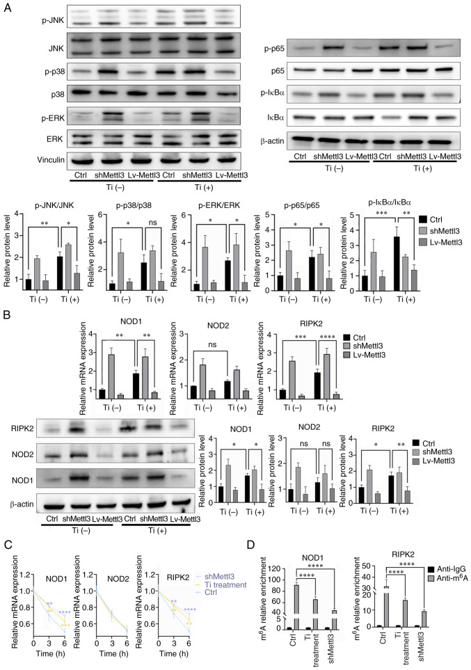
Peri‑prosthetic osteolysis (PPO) induced by wear particles is considered the primary cause of titanium prosthesis failure and revision surgery. The specific molecular mechanisms involve titanium particles inducing multiple intracellular pathways, which impact disease prevention and the targeted therapy of PPO. Notably, N6‑methyladenosine (m6A) serves critical roles in epigenetic regulation, particularly in bone metabolism and inflammatory responses. Thus, the present study aimed to determine the role of RNA methylation in titanium particle‑induced osteolysis. Results of reverse transcription‑quantitative PCR (RT‑qPCR), western blotting, ELISA and RNA dot blot assays revealed that titanium particles induced osteogenic inhibition and proinflammatory responses, accompanied by the reduced expression of methyltransferase‑like (Mettl) 3, a key component of m6A methyltransferase. Specific lentiviruses vectors were employed for Mettl3 knockdown and overexpression experiments. RT‑qPCR, western blotting and ELISA revealed that the knockdown of Mettl3 induced osteogenic inhibition and proinflammatory responses comparable with that induced by titanium particle, while Mettl3 overexpression attenuated titanium particle‑induced cellular reactions. Methylated RNA immunoprecipitation‑qPCR results revealed that titanium particles mediated the methylation of two inhibitory molecules, namely Smad7 and SMAD specific E3 ubiquitin protein ligase 1, via Mettl3 in bone morphogenetic protein signaling, leading to osteogenic inhibition. Furthermore, titanium particles induced activation of the nucleotide binding oligomerization domain 1 signaling pathway through methylation regulation, and the subsequent activation of the MAPK and NF‑κB pathways. Collectively, the results of the present study indicated that titanium particles utilized Mettl3 as an upstream regulatory molecule to induce osteogenic inhibition and inflammatory responses. Thus, the present study may provide novel insights into potential therapeutic targets for aseptic loosening in titanium prostheses.

Keywords: N6‑methyladenosine RNA methylation; aseptic loosening; epigenetic regulation; methyltransferase‑like 3; osteolysis; titanium particles.


Conflict of interest statement

The authors declare that they have no competing interests.

Figures

Figure 1.
Figure 1.
Titanium particles inhibit osteogenic differentiation and mediate osteoblast-osteoclast communication. (A) Transmission electron microscopy of osteoblasts co-cultured (A-a) without and (A-b) with titanium particles (red arrows; scale bar, 5 µm). (B) Titanium particle treatment inhibited the formation of mineralized nodules at 14 days. (B-a) Blank, (B-b) osteogenic induction medium and (B-c) titanium particle treatment. Scale bar, 500 µm. (C) Titanium particle treatment inhibited the expression of osteogenesis-associated markers. The samples were collected at 2 and 3 days for RT-qPCR and western blotting, respectively. (D) Titanium particle treatment promoted the expression of inflammatory cytokines. The samples were collected at 2 and 3 days for RT-qPCR and ELISA, respectively. (E) Supernatants of titanium-treated osteoblasts promoted the expression of osteoclast differentiation-associated markers in preosteoclasts. (F) Titanium treatment promoted the expression of RANKL, leading to the higher ratio of RANKL/OPG. The samples were collected at 2 and 3 days for RT-qPCR and ELISA, respectively. Data are representative of three independent experiments and are presented as the mean ± standard deviation. *P<0.05, **P<0.01, ***P<0.001 and ****P<0.0001. Acp5, acid phosphatase 5, tartrate resistant; Col1a1, collagen type I α1 chain; Ctsk, cathepsin K; CM, conditioned medium; Dcstamp, dendrocyte expressed seven transmembrane protein; M-CSF, macrophage colony-stimulating factor; NC, negative control; Nfatc1, nuclear factor of activated T cells 1; ns, not significant; OD, optical density; OM, osteogenic induction medium; OPG, osteoprotegerin; PC, positive control; RANKL, receptor activator of NF-κB ligand; RT-qPCR, reverse transcription-quantitative PCR; Runx2, RUNX family transcription factor 2; Ti, titanium particle.
Figure 2.
Figure 2.
Role of m6A modification in titanium particle treatment. (A) Titanium particle treatment reduced total m6A levels compared with the osteogenic induction group after 2 days. (B) Titanium particle treatment significantly reduced the mRNA expression of the methylation-associated enzyme Mettl3. Samples were collected at 2 days for reverse transcription-quantitative PCR. (C) Titanium particle treatment significantly reduced the protein expression levels of Mettl3. Samples were collected at 3 days for western blotting. Data are representative of three independent experiments and are presented as the mean ± standard deviation. *P<0.05, **P<0.01 and ***P<0.001. Alkbh5, alkB homolog 5, RNA demethylase; Fto, FTO α-ketoglutarate dependent dioxygenase; m6A, N6-methyladenosine; Mettl3, methyltransferase-like 3; ns, not significant; OM, osteogenic induction medium; Ti, titanium particle.
Figure 3.
Figure 3.
Mettl3 knockdown induces osteogenic inhibition and proinflammatory responses. (A) Construction of Mettl3 knockdown cells (scale bar, 100 µm). (B) The verification of transfection efficiency using reverse transcription-quantitative PCR and western blotting. (C) Mettl3 knockdown and titanium particle treatment were associated with reduced m6A content in total RNA compared with the blank group (osteoblasts without transfection). (D) Mettl3 knockdown inhibited the expression of osteogenic markers and the formation of mineralized nodules (scale bar, 500 µm). (E) Mettl3 knockdown promoted the expression of inflammatory cytokines. (F) Mettl3 knockdown led to a higher ratio of RANKL/OPG compared with that of the control group (shCtrl cells without Ti). (G) Supernatant of Mettl3 knockdown osteoblasts promoted the expression of osteoclast differentiation-associated markers in preosteoclasts. Data are representative of three independent experiments and are presented as the mean ± standard deviation. *P<0.05, **P<0.01, ***P<0.001 and ****P<0.0001. Acp5, acid phosphatase 5, tartrate resistant; Col1a1, collagen type I α1 chain; Ctrl, control; Ctsk, cathepsin K; Dcstamp, dendrocyte expressed seven transmembrane protein; m6A, N6-methyladenosine; Mettl3, methyltransferase-like 3; Nfatc1, nuclear factor of activated T cells 1; ns, not significant; OD, optical density; OPG, osteoprotegerin; RANKL, receptor activator of NF-κB ligand; sh, short hairpin RNA; Runx2, RUNX family transcription factor 2; Ti, titanium particle.
Figure 4.
Figure 4.
Mettl3 overexpression attenuates titanium particle-induced osteogenesis inhibition and proinflammatory responses. (A) Construction of Mettl3 overexpression cells and the verification of transfection efficiency using reverse transcription-quantitative PCR and western blotting (scale bar, 100 µm). (B) Transmission electron microscopy of Mettl3 overexpression cells co-cultured (B-a) without or (B-b) with titanium particles (red arrows; scale bar, 5 µm). (C) Mettl3 overexpression increased the m6A content in total RNA compared with that of the blank group (osteoblasts without transfection). (D) Mettl3 overexpression attenuated titanium particle-induced osteogenesis inhibition (scale bar, 500 µm). (E) Mettl3 overexpression attenuated titanium particle-induced proinflammatory responses. (F) Mettl3 overexpression attenuated the titanium particle-induced increase in the RANKL/OPG ratio. (G) Mettl3 overexpression attenuated osteoclast differentiation promotion induced by the supernatant of Ti-treated osteoblasts. Data are representative of three independent experiments and are presented as the mean ± standard deviation. *P<0.05, **P<0.01, ***P<0.001 and ****P<0.0001. Acp5, acid phosphatase 5, tartrate resistant; Col1a1, collagen type I α1 chain; Ctsk, cathepsin K; Dcstamp, dendrocyte expressed seven transmembrane protein; Lv, lentivirus; m6A, N6-methyladenosine; Mettl3, methyltransferase-like 3; Nfatc1, nuclear factor of activated T cells 1; ns, not significant; OD, optical density; OPG, osteoprotegerin; RANKL, receptor activator of NF-kB ligand; Runx2, RUNX family transcription factor 2; Ti, titanium particle.
Figure 5.
Figure 5.
Titanium particle treatment targets Smad7 and Smurf1 via Mettl3, leading to BMP signaling inhibition. (A) Effects of titanium particle treatment and Mettl3 knockdown/overexpression on the mRNA expression of key signaling molecules of BMP signaling. (B) Effects of titanium particle treatment and Mettl3 knockdown/overexpression on BMP signaling activation. (C) Titanium particle treatment enhanced the mRNA stability of Smad7 and Smurf1 transcripts at the selected timepoints, following the addition of actinomycin D. (D) Titanium particle treatment significantly decreased the m6A modification of these two transcripts. The control group in this figure were osteoblasts without transfection. Data are representative of three independent experiments and are presented as the mean ± standard deviation. *P<0.05, **P<0.01, ***P<0.001 and ****P<0.0001. BMP, bone morphogenetic protein; Ctrl, control; Lv, lentivirus; m6A, N6-methyladenosine; Mettl3, methyltransferase-like 3; ns, not significant; p-, phosphorylated; sh, short hairpin RNA; Smurf1, SMAD specific E3 ubiquitin protein ligase 1; t-, total; Ti, titanium particle.
Figure 6.
Figure 6.
Titanium particle treatment induces NOD-like receptors to exert proinflammatory responses. (A) Titanium particle treatment induced the activation of the MAPK and NF-κB signaling pathways. (B) Titanium particle treatment induced the activation of the NOD-like receptor pathway, while Mettl3 overexpression attenuated these effects. (C) Titanium particle treatment and Mettl3 knockdown enhanced the mRNA stabilities of NOD1 and RIPK2 following the addition of actinomycin D. (D) Titanium particle treatment and Mettl3 knockdown decreased the m6A modification of NOD1 and RIPK2. The control group in this figure were osteoblasts without transfection. Data are representative of three independent experiments and are presented as the mean ± standard deviation. *P<0.05, **P<0.01, ***P<0.001 and ****P<0.0001. Ctrl, control; Lv, lentivirus; m6A, N6-methyladenosine; Mettl3, methyltransferase-like 3; NOD, nucleotide binding oligomerization domain; ns, not significant; p-, phosphorylated; RIPK2, receptor interacting serine/threonine kinase 2; sh, short hairpin RNA; Ti, titanium particle.
Figure 7.
Figure 7.
Inhibition of the NOD-like receptor signaling pathway inhibits titanium particle-induced proinflammatory responses. (A) Inhibition of the NOD1 signaling pathway inhibited MAPK and NF-κB signaling activation induced by titanium particle treatment. (B) Inhibition of the NOD1 signaling pathway inhibited proinflammatory cytokine expression induced by titanium particle treatment. The control group in this figure were osteoblasts without inhibitors. Data are representative of three independent experiments and are presented as the mean ± standard deviation. *P<0.05, **P<0.01 and ***P<0.001. Ctrl, control; Mettl3, methyltransferase-like 3; NOD, nucleotide binding oligomerization domain; p-, phosphorylated; sh, short hairpin RNA; Ti, titanium particle.
Figure 8.
Figure 8.
Ythdf2 participates in the methyltransferase-like 3-mediated bioactivities in titanium particle treatment. (A) Knockdown of Ythdf2 and verification of transfection efficiency using reverse transcription-quantitative PCR and western blotting. (B) Ythdf2 knockdown promoted the expression of Smad7, Smurf1, NOD1 and RIPK2. (C) Ythdf2 knockdown enhanced the mRNA stabilities of Smad7, Smurf1, NOD1 and RIPK2 at 3 h. (D) Methylated RNA immunoprecipitation-quantitative PCR results demonstrated that titanium particle treatment decreased the relative expression levels of Smad7, Smurf1, NOD1 and RIPK2 precipitated by Ythdf2. Data are representative of three independent experiments and are presented as the mean ± standard deviation. *P<0.05, **P<0.01, ***P<0.001 and ****P<0.0001. Ctrl, control; NOD, nucleotide binding oligomerization domain; RIPK2, receptor interacting serine/threonine kinase 2; si, small interfering RNA; Smurf1, SMAD specific E3 ubiquitin protein ligase 1; Ythdf2, YTH domain family 2; Ti, titanium particle.

Similar articles

Cited by

References

    1. Ollivere B, Wimhurst JA, Clark IM, Donell ST. Current concepts in osteolysis. J Bone Joint Surg Br. 2012;94:10–15. doi: 10.1302/0301-620X.94B1.28047. - DOI - PubMed
    1. Hodges NA, Sussman EM, Stegemann JP. Aseptic and septic prosthetic joint loosening: Impact of biomaterial wear on immune cell function, inflammation, and infection. Biomaterials. 2021;278:121127. doi: 10.1016/j.biomaterials.2021.121127. - DOI - PubMed
    1. Tay ML, Matthews BG, Monk AP, Young SW. Disease progression, aseptic loosening and bearing dislocations are the main revision indications after lateral unicompartmental knee arthroplasty: A systematic review. J ISAKOS. 2022;7:132–141. doi: 10.1016/j.jisako.2022.06.001. - DOI - PubMed
    1. Eger M, Sterer N, Liron T, Kohavi D, Gabet Y. Scaling of titanium implants entrains inflammation-induced osteolysis. Sci Rep. 2017;7:39612. doi: 10.1038/srep39612. - DOI - PMC - PubMed
    1. McArthur BA, Scully R, Patrick Ross F, Bostrom MPG, Falghren A. Mechanically induced periprosthetic osteolysis: A systematic review. HSS J. 2019;15:286–296. doi: 10.1007/s11420-018-9641-5. - DOI - PMC - PubMed