. 2024 Feb 20;9(2):e0057223.
doi: 10.1128/msystems.00572-23. Epub 2024 Jan 12.

Periodontitis aggravates COPD through the activation of γδ T cell and M2 macrophage

Affiliations

Periodontitis aggravates COPD through the activation of γδ T cell and M2 macrophage

Kaixin Xiong et al. mSystems. .

Abstract

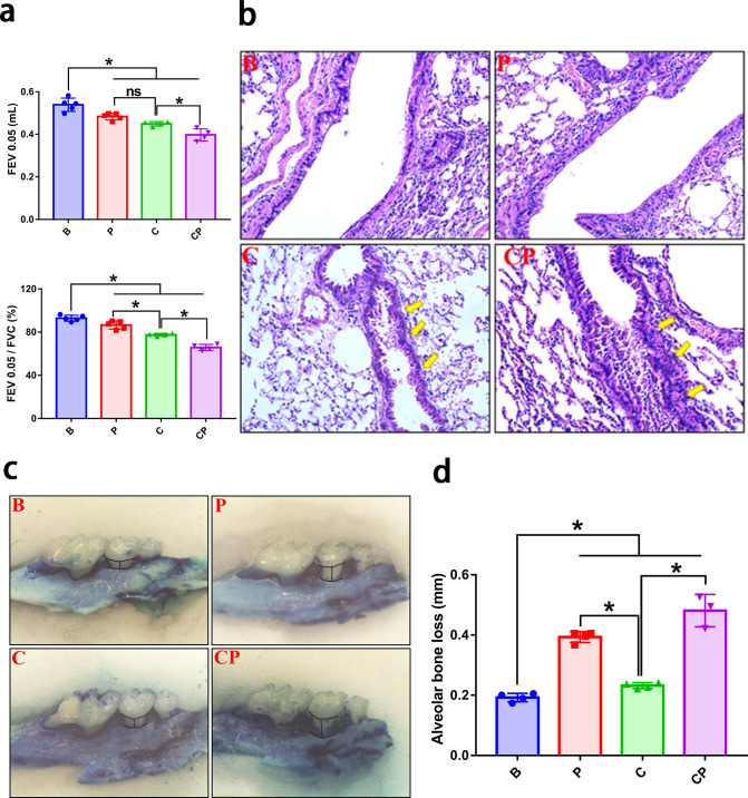
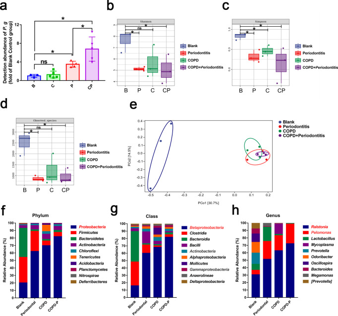
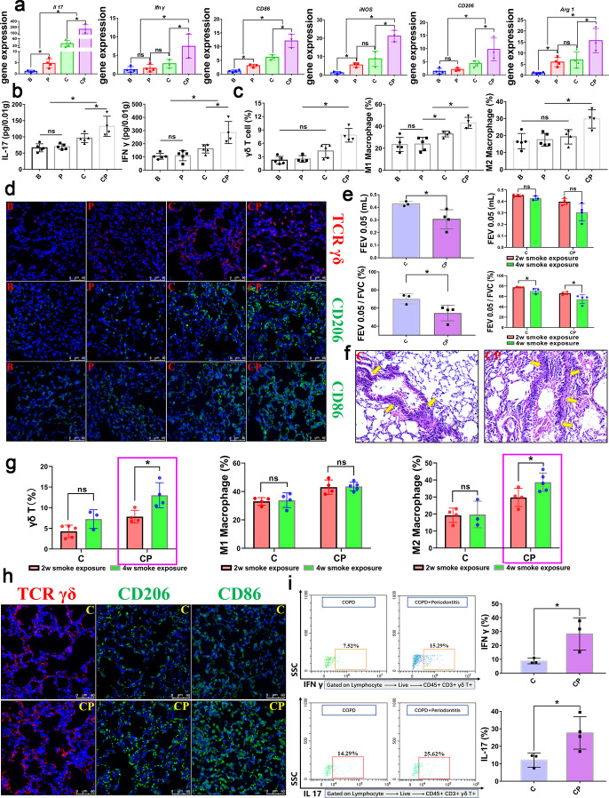
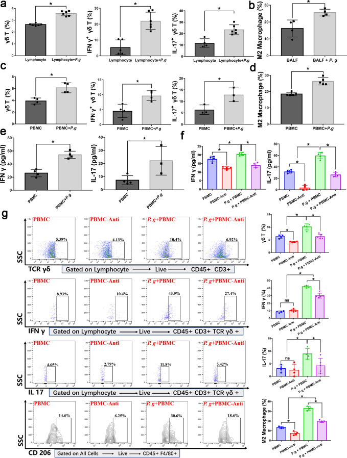
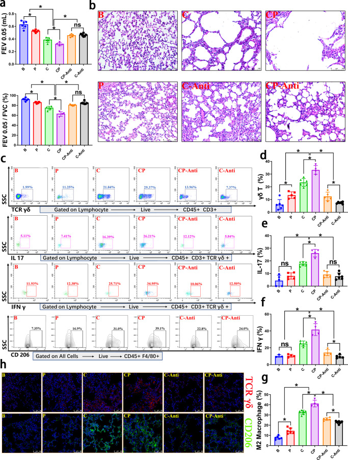
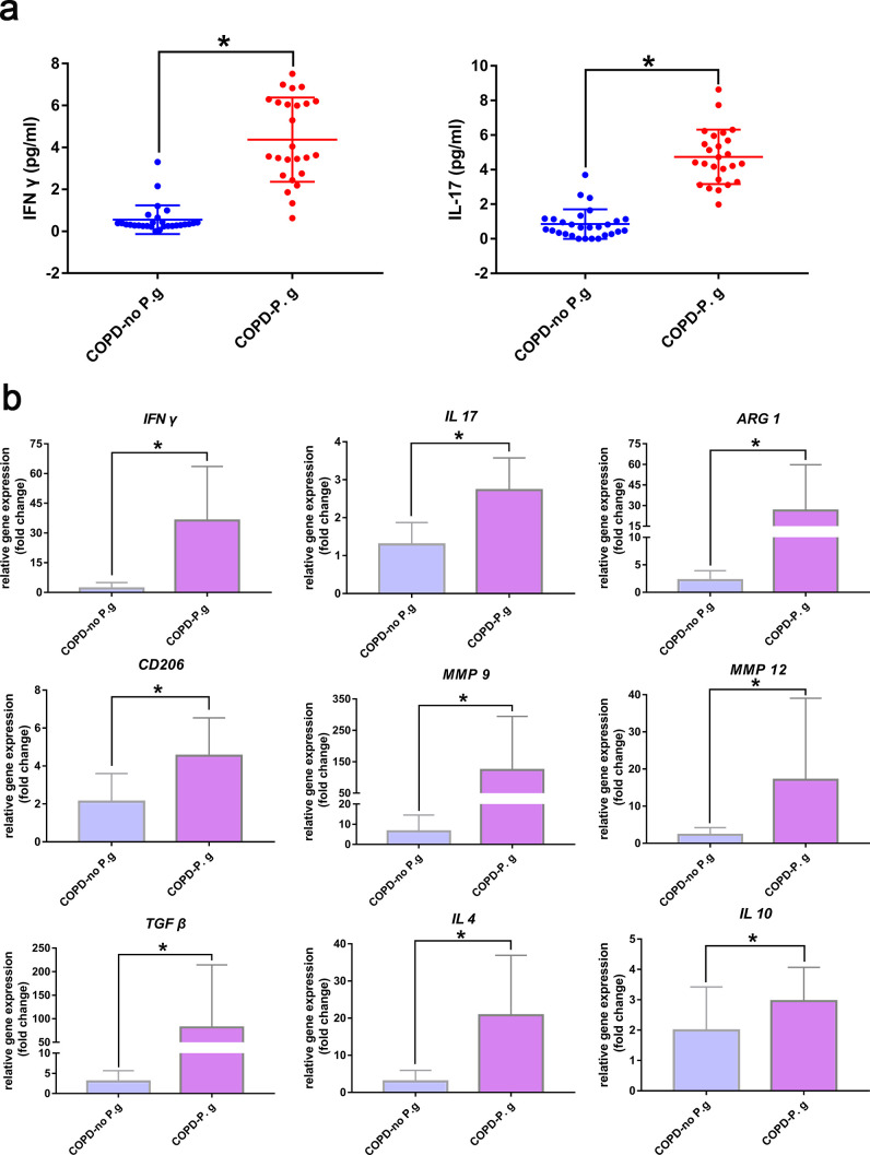
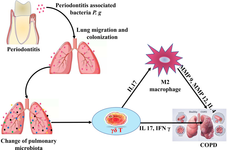
Chronic obstructive pulmonary disease (COPD) is a chronic systemic inflammatory disease with high morbidity and mortality. Periodontitis exacerbates COPD progression; however, the immune mechanisms by which periodontitis affects COPD remain unclear. Here, by constructing periodontitis and COPD mouse models, we demonstrated that periodontitis and COPD could mutually aggravate disease progression. For the first time, we found that the progression was associated with the activation of γδ T cells and M2 macrophages, and M2 polarization of macrophages was affected by γδ T cells activation. In the lung tissues of COPD with periodontitis, the activation of γδ T cells finally led to the increase of IL 17 and IFN γ expression and M2 macrophage polarization. Furthermore, we found that the periodontitis-associated bacteria Porphyromonas gingivalis (P. gingivalis) promoted the activation of γδ T cells and M2 macrophages ex vivo. The data from clinical bronchoalveolar lavage fluid (BALF) samples were consistent with the in vivo and ex vivo experiments. For the first time, our results identified the crucial role of γδ T-M2 immune mechanism in mediating periodontitis-promoted COPD progression. Therefore, targeting at periodontitis treatment and the γδ T-M2 immune mechanism might provide a new practical strategy for COPD prevention or control.IMPORTANCEPeriodontitis exacerbates chronic obstructive pulmonary disease (COPD) progression. For the first time, the current study identified that the impact of periodontitis on COPD progression was associated with the activation of γδ T cells and M2 macrophages and that M2 polarization of macrophages was affected by γδ T cells activation. The results indicated that targeting at periodontitis treatment and the γδ T-M2 immune mechanism might provide a new practical strategy for COPD prevention or control.

Keywords: COPD; IFN γ; IL 17; M2 macrophage; periodontitis; γδ T.


Conflict of interest statement

The authors declare no conflict of interest.

Figures

Fig 1
Fig 1
Periodontitis and COPD mutually promoted disease progression. (a) The lung function test was carried out to assess the severity of COPD, and the results were presented with FEV0.05 value and FEV0.05/FVC value for each group. FEV0.05: forced expiratory volume in 0.05 s. FEV0.05/FVC: ratio of forced expiratory volume in 0.05 s to forced vital capacity. (b) H&E stain was performed to observe the lung morphology change and a representative H&E image of lung tissue was shown for each group. (c) The methylene blue stain was carried out to analyze the periodontitis alveolar bone loss and a representative image of the jaw was shown for each group. (d) Quantitative analysis of alveolar bone loss in each group. *P < 0.05. B, blank control; P, periodontitis; C, COPD; CP, COPD with periodontitis.
Fig 2
Fig 2
P. gingivalis-periodontitis affected homeostasis of the lung microbiota. (a) The relative abundance of P. gingivalis in lung tissues was determined by RT-qPCR. (b–d) α-diversity of the lung tissue was analyzed for each group and presented with Shannon, Simpson, and observed species indices. (e) β diversity of the lung tissue in each group was presented with PCOA index. (f–h) Lung microbial community composition was analyzed. The percentages of the major phyla, classes, and genera according to 16S rRNA sequencing were shown. *P < 0.05, ns, not significant; B, blank control; P, periodontitis; C, COPD; CP, COPD with periodontitis.
Fig 3
Fig 3
Periodontitis exacerbated COPD through promoting γδ T cells aggregation and macrophages polarization in the lung tissue, and the promotive effect was prolonged over time. (a–d) Results of the early stage of COPD (short smoke exposure time, 2 weeks of daily exposure) model; (e–i) Results of the late stage of COPD (extended smoke exposure time, 4 weeks of daily exposure) model. (a) In the early stage of COPD, the relative mRNA expression of γδ T-related genes, Il 17 and Ifn γ, M1-related genes, CD86 and iNOS, M2-related genes, CD206 and Arg 1 in the lung tissue of each group were determined by RT-qPCR and displayed with the fold change to B group. (b) The levels of IL 17 and IFN γ in lung tissue of each group were calculated by ELISA for early stage of COPD. (c, d) In the early stage of COPD, the percentages of γδ T positive cells, M1 polarized macrophages, and M2 polarized macrophages in the lung tissue of each group were quantified by flow cytometry analysis (γδ T cells were gated on the lymphocytes, live cells, and CD45 positive and CD3 positive cells. M1 and M2 cells were gated on all cells, live cell, and CD45 positive and F4/80 positive cells.) and observed by confocal immunofluorescence analysis. Blue color, DAPI; red color, TCR γδ; green color, CD206 or CD86. Following, we further extended the smoke exposure time to 4 weeks and analyzed the effect of periodontitis on COPD in the time-extended (late-stage) model. (e) In the late stage of COPD, the lung function test results of each group and comparison with the corresponding early stage of COPD (2 weeks smoke exposure) were analyzed. FEV0.05, forced expiratory volume in 0.05 s. FEV0.05/FVC: ratio of forced expiratory volume in 0.05 s to forced vital capacity. (f) Representative H&E images of lung tissues in the late stage of COPD (4 weeks smoke exposure) were shown. (g) The quantitative cell numbers calculated by flow cytometry between early stage and late stage of COPD were compared, among which γδ T cells and M2 macrophages showed a significant increase, so, in the following research, we focused on these two types of immune cells. (h) Representative confocal immunofluorescence images of lung tissue in the late stage of COPD. Blue color, DAPI; red color, TCR γδ; green color, CD206 or CD86. (i) In the late stage of COPD, we further observed periodontitis promoted the aggregation of IL 17+ γδ T cells and IFN γ+ γδ T cells. Representative flow cytometry plots of IL 17+ γδ T cells and IFN γ+ γδ T cells and corresponding quantitative analysis of lung tissue in late stage of COPD were shown. *P < 0.05, ns, not significant. B, blank control; P, periodontitis; C, COPD; CP, COPD with periodontitis.
Fig 4
Fig 4
Periodontitis-associated bacteria P. gingivalis promoted γδ T cells activation and M2 polarization in the lung tissue and PBMC. The promotion effects were affected by γδ-TCR monoclonal antibody treatment. These ex vivo experiments were conducted using mice materials, including mice lung tissue lymphocytes, BALF and PBMC. (a, b) P. gingivalis promoted γδ T cells activation in lung tissue lymphocytes, and M2 macrophages in the BALF, the percentages of γδ T cells, IFN γ+ γδ T cells, IL 17+ γδ T cells (a) and M2 macrophages (b) were calculated. (c, d) P. gingivalis promoted γδ T cells activation and M2 polarization in the PBMC, the percentages of γδ T cells, IFN γ+ γδ T cells, IL 17+ γδ T cells (c) and M2 macrophages (d) were calculated. (e, f) P. gingivalis promoted the levels of IL 17 and IFN γ in the PBMC supernatants (e), and the promotion effect was impacted by γδ-TCR monoclonal antibody treatment (f). (g) With or without γδ-TCR monoclonal antibody treatment, P. gingivalis were cocultured with PBMC. γδ T cells activation and M2 polarization were analyzed by flow cytometry and representative flow cytometry plots were shown, and the corresponding quantitative analysis of γδ T cells, IL 17+ γδ T cells, IFN γ+ γδ T cells, and M2 cells was presented as mean ± STD. *P < 0.05, ns, not significant. Lung-lym, the lymphocytes of lung tissue; Lung-lym +P. g, the lymphocytes of lung tissue cocultured with P. gingivalis; BALF, bronchoalveolar lavage fluid; BALF +P. g, bronchoalveolar lavage fluid cocultured with P. gingivalis; PBMC, peripheral blood mononuclear cells; PBMC +P. g, peripheral blood mononuclear cells cocultured with P. gingivalis; PBMC-Anti, PBMC group with γδ-TCR monoclonal antibody treatment; P. g + PBMC-Anti, P. g + PBMC group with γδ-TCR monoclonal antibody treatment.
Fig 5
Fig 5
Inhibition of γδ T cells affected periodontitis promoting COPD in vivo. (a) With or without the γδ-TCR monoclonal antibody treatment, the lung function test was performed in each group, and the results were shown as FEV0.05 value and FEV0.05/FVC value. FEV0.05, forced expiratory volume in 0.05 s. FEV0.05/FVC, ratio of forced expiratory volume in 0.05 s to forced vital capacity. (b) A representative H&E image of lung tissue in each group was shown. Periodontitis aggravated COPD, while γδ-TCR monoclonal antibody treatment partly reduced COPD severity. (c–g): Representative flow cytometry plots and corresponding quantitative analysis of lung tissue for each group were shown. (h) With or without γδ-TCR monoclonal antibody treatment, representative confocal immunofluorescence images of γδ T cells and M2 macrophages of lung tissue for each group were shown. Blue color, DAPI; red color, TCR γδ; green color, CD206. *P < 0.05, ns, not significant. B, blank control; P, periodontitis; C, COPD; CP, COPD with periodontitis; C-Anti, the COPD group with γδ-TCR monoclonal antibody treatment; CP-Anti, the CP group with γδ-TCR monoclonal antibody treatment.
Fig 6
Fig 6
Enhanced IFN γ and IL 17 levels and expression of M2-polarized genes in clinical COPD with periodontal pathogen BALF samples. (a) IFN γ and IL 17 levels in clinical COPD BALF samples were measured by ELISA. (b) Relative gene expression levels in clinical COPD BALF samples were quantified by RT-qPCR. *P < 0.05, ns, not significant. COPD-no P. g, BALF of COPD patients with no P. gingivalis detected; COPD-P. g, BALF of COPD patients with P. gingivalis detected.
Fig 7
Fig 7
The potential immune mechanism by which periodontitis aggravates COPD progression. Under periodontitis state, periodontitis-associated bacteria, as represented by P. gingivalis, migrate to and colonize on lung tissue and affect lung microbiota homeostasis. Following, periodontitis promotes the aggregation and activation of γδ T cells, with IL 17 and IFN γ directly promoting the progression of COPD. Simultaneously, IL 17 further induces M2 polarization of macrophages, thereby contributing to COPD progression.

Similar articles

Cited by

References

    1. Moghadam SA, Shirzaiy M, Risbaf S. 2017. The associations between periodontitis and respiratory disease. J Nepal Health Res Counc 15:1–6. doi:10.3126/jnhrc.v15i1.18023 - DOI - PubMed
    1. Pathak JL, Yan Y, Zhang Q, Wang L, Ge L. 2021. The role of oral microbiome in respiratory health and diseases. Respir Med 185:106475. doi:10.1016/j.rmed.2021.106475 - DOI - PubMed
    1. Ahmad Hassali MA, Muhammad SA, Shah S, Abbas S, Hyder Ali IAB, Salman A. 2020. The economic burden of chronic obstructive pulmonary disease (COPD) in the USA, Europe, and Asia: results from a systematic review of the literature. Expert Rev Pharmacoecon Outcomes Res 20:661–672. doi:10.1080/14737167.2020.1678385 - DOI - PubMed
    1. Lortet-Tieulent J, Soerjomataram I, López-Campos JL, Ancochea J, Coebergh JW, Soriano JB. 2019. International trends in COPD mortality. Eur Respir J 54:1995–2017. doi:10.1183/13993003.01791-2019 - DOI - PubMed
    1. Hajishengallis G, Chavakis T. 2021. Local and systemic mechanisms linking periodontal disease and inflammatory comorbidities. Nat Rev Immunol 21:426–440. doi:10.1038/s41577-020-00488-6 - DOI - PMC - PubMed