. 2024 Feb 20;98(2):e0137223.
doi: 10.1128/jvi.01372-23. Epub 2024 Jan 12.

Prefusion stabilization of the Hendra and Langya virus F proteins

Affiliations

Prefusion stabilization of the Hendra and Langya virus F proteins

Patrick O Byrne et al. J Virol. .

Abstract

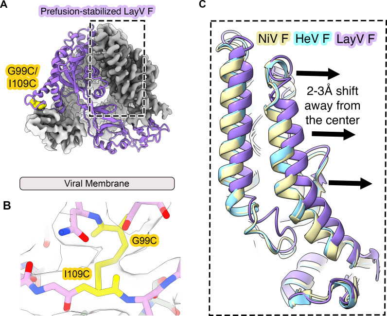
Nipah virus (NiV) and Hendra virus (HeV) are pathogenic paramyxoviruses that cause mild-to-severe disease in humans. As members of the Henipavirus genus, NiV and HeV use an attachment (G) glycoprotein and a class I fusion (F) glycoprotein to invade host cells. The F protein rearranges from a metastable prefusion form to an extended postfusion form to facilitate host cell entry. Prefusion NiV F elicits higher neutralizing antibody titers than postfusion NiV F, indicating that stabilization of prefusion F may aid vaccine development. A combination of amino acid substitutions (L104C/I114C, L172F, and S191P) is known to stabilize NiV F in its prefusion conformation, although the extent to which substitutions transfer to other henipavirus F proteins is not known. Here, we perform biophysical and structural studies to investigate the mechanism of prefusion stabilization in F proteins from three henipaviruses: NiV, HeV, and Langya virus (LayV). Three known stabilizing substitutions from NiV F transfer to HeV F and exert similar structural and functional effects. One engineered disulfide bond, located near the fusion peptide, is sufficient to stabilize the prefusion conformations of both HeV F and LayV F. Although LayV F shares low overall sequence identity with NiV F and HeV F, the region around the fusion peptide exhibits high sequence conservation across all henipaviruses. Our findings indicate that substitutions targeting this site of conformational change might be applicable to prefusion stabilization of other henipavirus F proteins and support the use of NiV as a prototypical pathogen for henipavirus vaccine antigen design.IMPORTANCEPathogenic henipaviruses such as Nipah virus (NiV) and Hendra virus (HeV) cause respiratory symptoms, with severe cases resulting in encephalitis, seizures, and coma. The work described here shows that the NiV and HeV fusion (F) proteins share common structural features with the F protein from an emerging henipavirus, Langya virus (LayV). Sequence alignment alone was sufficient to predict which known prefusion-stabilizing amino acid substitutions from NiV F would stabilize the prefusion conformations of HeV F and LayV F. This work also reveals an unexpected oligomeric interface shared by prefusion HeV F and NiV F. Together, these advances lay a foundation for future antigen design targeting henipavirus F proteins. In this way, Nipah virus can serve as a prototypical pathogen for the development of protective vaccines and monoclonal antibodies to prepare for potential henipavirus outbreaks.

Keywords: Hendra; Langya; Nipah; electron microscopy; paramyxovirus; prefusion; structure-based vaccine design.


Conflict of interest statement

J.S.M., R.J.L., and B.S.G. are inventors on U.S. patent application no. 62/714,230 ("Nipah virus Immunogens and Their Use"). B.S.G. and R.J.L. are inventors on US patent application no. 63/315,934 (filed 2 March 2022, "Monoclonal Antibodies to Nipah Virus F Protein and Their Use").

Figures

Fig 1
Fig 1
Known prefusion-stabilizing substitutions from NiV F target three sites of predicted conformational change. Two ribbon diagrams of individual protomers of prefusion (far left, pale rainbow) and postfusion (far right, bright rainbow) NiV F. The N-terminal ends are colored blue, the C-terminal ends are colored red. (A–D) Circled letters indicate areas of interest and correspond to zoomed views in panels A, B, C, and D. Arrows in panels A–D indicate broad conformational movements from prefusion (pale colors) to postfusion (bright colors). In some panels, the movement to postfusion is out of frame and is indicated by a distance or angle estimate. (E) Multiple sequence alignment of F proteins from nine henipaviruses: Henipavirus nipahense (Malaysia strain, AAK29087.1), Henipavirus nipahense (Bangladesh strain, AAY43915.1), Henipavirus hendraense (NP_047111.2), Henipavirus hendraense (g2 strain, UCY33687.1), Henipavirus mòjiāngense (YP_009094094.1), Henipavirus cedarense (YP_009094085.1), Henipavirus ghanaense (YP_009091837.1), Langya virus (UUV47240.1), and Angavokely virus (UVG43988.1). The region near the fusion peptide and cleavage site is colored with a light red box. Sites of stabilizing substitutions in NiV F are indicated with colored arrows: disulfide (yellow), cavity-filling (purple), and proline (green). Sequence conservation is colored according to the legend at the bottom of (E).
Fig 2
Fig 2
Purification and characterization of prefusion-stabilized Hendra virus F protein. (A–B) SEC of wild-type HeV F, single variants and the triple variant. UV traces for disulfide, cavity-filling, and proline substitutions are colored orange, purple, and green, respectively. The dotted line extending from (A) into (B) shows the position of high-molecular-weight peaks for the single variants, which disappear when all three substitutions are combined. Trimeric HeV F and NiV F are shown as dashed red and black traces, respectively. (C) DSF of wild-type HeV F and variants, showing the change in fluorescence with respect to temperature (dF/dT) as a function of temperature (°C). The colors of the DSF traces are the same as in the SEC traces from panels A and B.
Fig 3
Fig 3
Cryo-EM structures of prefusion-stabilized NiV F and HeV F. (A–B) Cryo-EM maps and models of prefusion-stabilized NiV F (A) and HeV F (B). Two protomers are represented as light gray cryo-EM maps, and the third protomer is shown as a ribbon (yellow for NiV F, blue for HeV F). Atoms corresponding to the three substitutions are shown as colored spheres (L104C/V114C are gold, S191P is green, L172F is purple). The viral membrane is shown as a gray tube. (C–G) Zoomed views of models and cryo-EM maps near the sites of disulfide (C–D), cavity-filling (E–F), and proline (G–H) substitutions. The cryo-EM maps are shown as a transparent gray surface.
Fig 4
Fig 4
A disulfide substitution near the fusion peptide stabilizes prefusion LayV F. (A) SEC of LayV F wild-type (WT) and three variants. The UV traces are overlaid for wild-type (black), G99C/I109C (orange), S186P (green), and L237F (purple). (B–E) negative-stain EM analysis of LayV F, showing 2D class averages for (B) wild-type LayV F ectodomain and three variants: (C) L237F, (D) S186P, and (E) G99C/I109C. (F) DSF of LayV F showing the change in fluorescence with respect to temperature (dF/dT) as a function of temperature (°C). The LayV F WT, S186P, L237F, and G99C/I109C variants are shown as solid black, green, purple, and yellow traces, respectively. Controls for prefusion NiV F and prefusion HeV F are shown as dashed black and red traces, respectively.
Fig 5
Fig 5
Cryo-EM structure of prefusion-stabilized LayV F G99C/I109C. (A) Cryo-EM map and model of the disulfide-stabilized LayV F variant G99C/I109C. One protomer is shown as a purple ribbon diagram and the cryo-EM map is shown for the other two protomers. (B) Zoomed view of the LayV F model within a transparent gray cryo-EM map at the disulfide substitution site. The cysteines at positions 99 and 109 are colored yellow. (C) Overlay of the prefusion models of NiV F (pale yellow), HeV F (cyan), and LayV F G99C/I109C (purple). The direction of the outward shift of the LayV F helix relative to NiV F and HeV F is indicated with arrows.
Fig 6
Fig 6
Cryo-EM structures of HeV F and NiV F dimers-of-trimers. (A) Side and top views of the cryo-EM map of a HeV F dimer-of trimers. Individual HeV F protomers (F1, F2, and F3) are colored in shades of blue. (B) Side and top views of a NiV F dimer-of-trimers. NiV F protomers are colored yellow, blue, and red. (C) Interface between two NiV F protomers on adjacent trimers. Side chains of residues at the dimer-of-trimers interface are shown as sticks. Hydrogen bonds are shown as light blue dashes.

References

    1. Chua KB, Bellini WJ, Rota PA, Harcourt BH, Tamin A, Lam SK, Ksiazek TG, Rollin PE, Zaki SR, Shieh W, Goldsmith CS, Gubler DJ, Roehrig JT, Eaton B, Gould AR, Olson J, Field H, Daniels P, Ling AE, Peters CJ, Anderson LJ, Mahy BW. 2000. Nipah virus: a recently emergent deadly paramyxovirus. Science 288:1432–1435. doi:10.1126/science.288.5470.1432 - DOI - PubMed
    1. Chua KB, Goh KJ, Wong KT, Kamarulzaman A, Tan PS, Ksiazek TG, Zaki SR, Paul G, Lam SK, Tan CT. 1999. Fatal encephalitis due to Nipah virus among pig-farmers in Malaysia. Lancet 354:1257–1259. doi:10.1016/S0140-6736(99)04299-3 - DOI - PubMed
    1. Mohd Nor MN, Gan CH, Ong BL. 2000. Nipah virus infection of pigs in peninsular Malaysia. Rev Sci Tech 19:160–165. doi:10.20506/rst.19.1.1202 - DOI - PubMed
    1. Arankalle VA, Bandyopadhyay BT, Ramdasi AY, Jadi R, Patil DR, Rahman M, Majumdar M, Banerjee PS, Hati AK, Goswami RP, Neogi DK, Mishra AC. 2011. Genomic characterization of Nipah virus, West Bengal, India. Emerg Infect Dis 17:907–909. doi:10.3201/eid1705.100968 - DOI - PMC - PubMed
    1. Goh KJ, Tan CT, Chew NK, Tan PS, Kamarulzaman A, Sarji SA, Wong KT, Abdullah BJ, Chua KB, Lam SK. 2000. Clinical features of Nipah virus encephalitis among pig farmers in Malaysia. N Engl J Med 342:1229–1235. doi:10.1056/NEJM200004273421701 - DOI - PubMed

Publication types

Supplementary concepts