Review
. 2024 Jan 12;147(1):15.
doi: 10.1007/s00401-023-02669-8.

Complement and MHC patterns can provide the diagnostic framework for inflammatory neuromuscular diseases

Affiliations
Review

Complement and MHC patterns can provide the diagnostic framework for inflammatory neuromuscular diseases

Christopher Nelke et al. Acta Neuropathol. .

Abstract

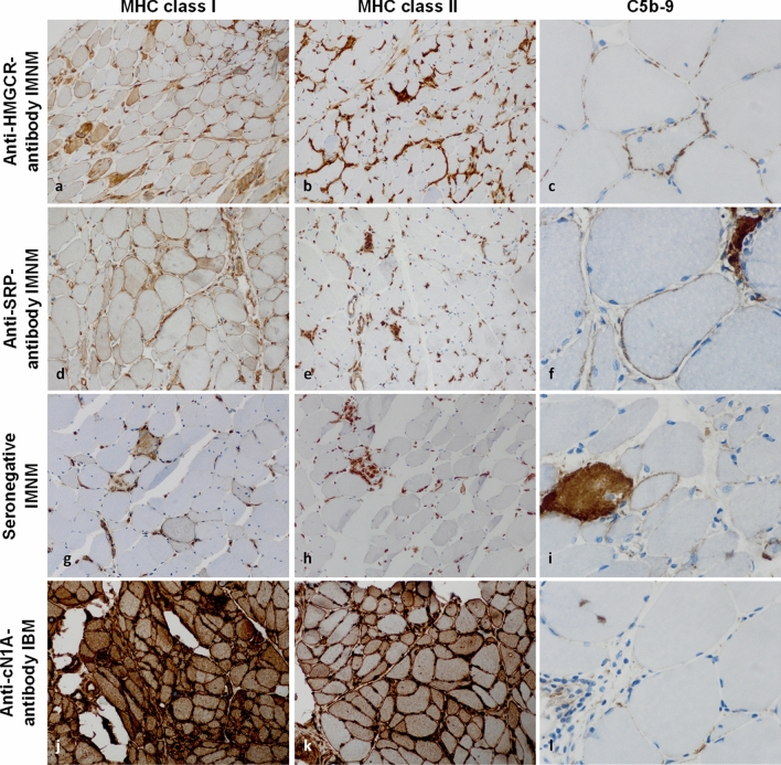
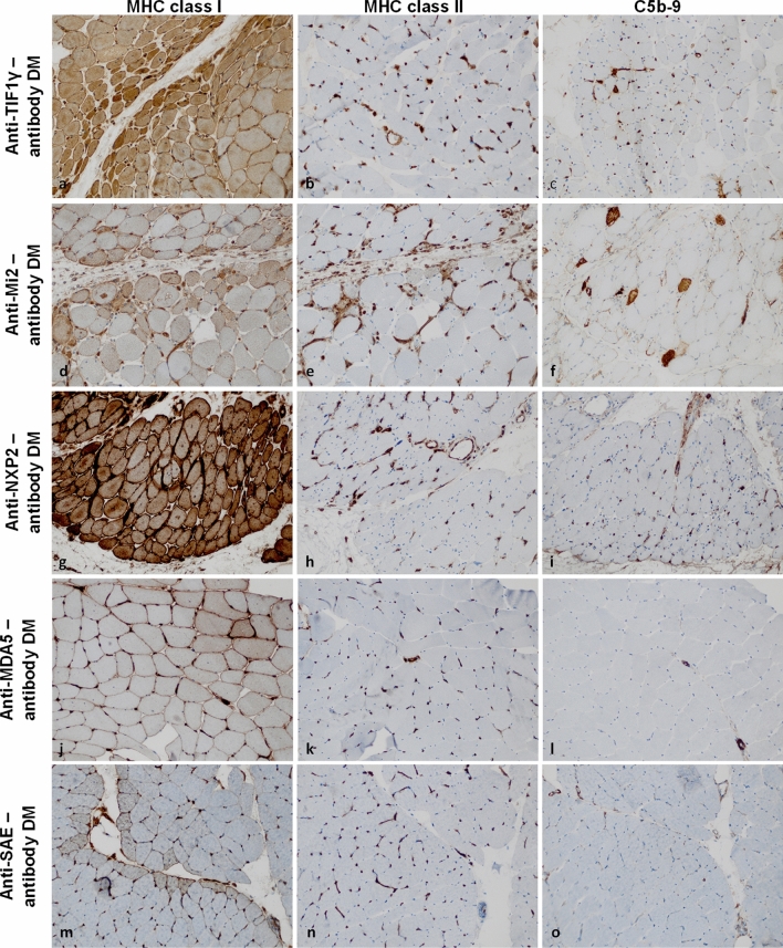
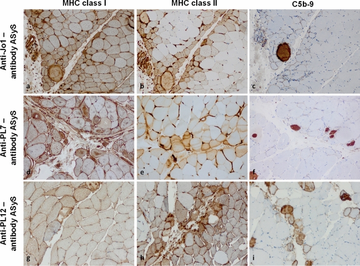
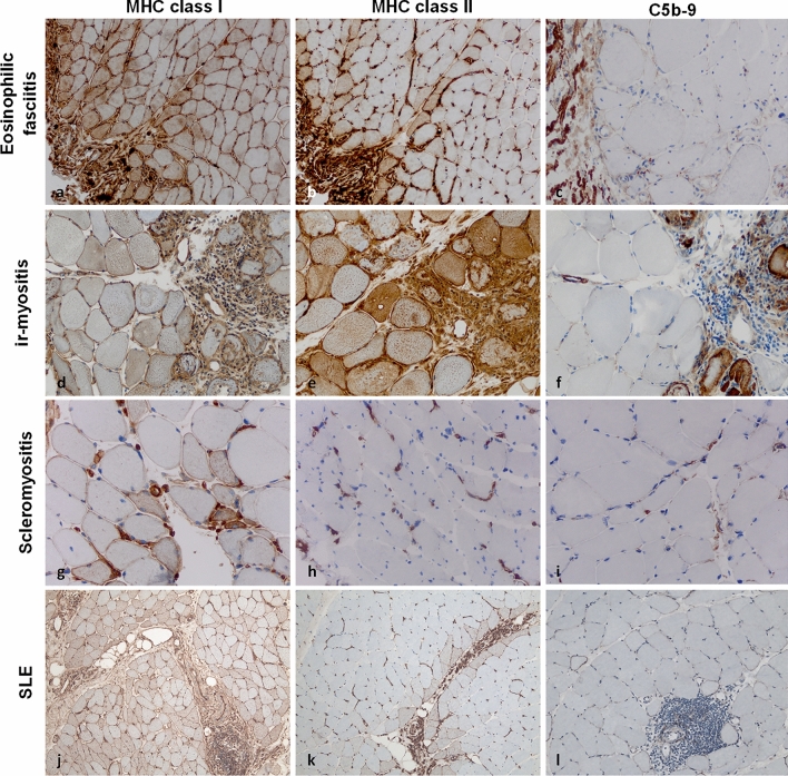
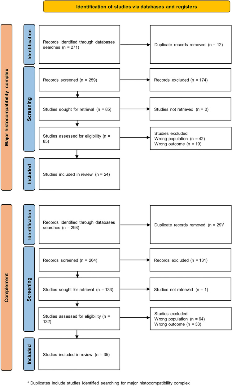
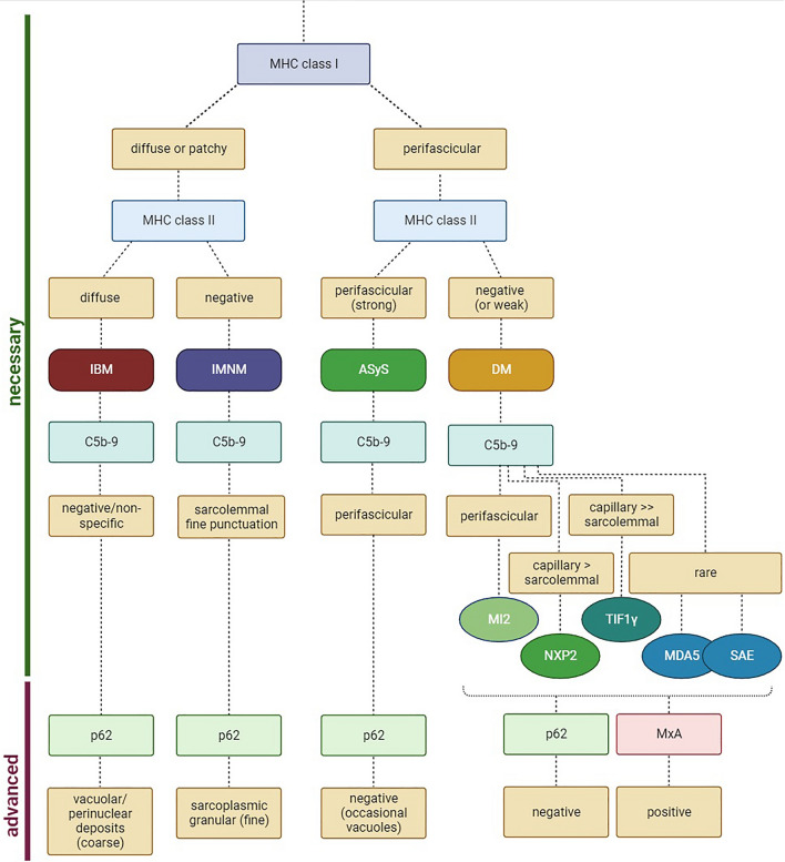
Histopathological analysis stands as the gold standard for the identification and differentiation of inflammatory neuromuscular diseases. These disorders continue to constitute a diagnostic challenge due to their clinical heterogeneity, rarity and overlapping features. To establish standardized protocols for the diagnosis of inflammatory neuromuscular diseases, the development of cost-effective and widely applicable tools is crucial, especially in settings constrained by limited resources. The focus of this review is to emphasize the diagnostic value of major histocompatibility complex (MHC) and complement patterns in the immunohistochemical analysis of these diseases. We explore the immunological background of MHC and complement signatures that characterize inflammatory features, with a specific focus on idiopathic inflammatory myopathies. With this approach, we aim to provide a diagnostic algorithm that may improve and simplify the diagnostic workup based on a limited panel of stainings. Our approach acknowledges the current limitations in the field of inflammatory neuromuscular diseases, particularly the scarcity of large-scale, prospective studies that validate the diagnostic potential of these markers. Further efforts are needed to establish a consensus on the diagnostic protocol to effectively distinguish these diseases.

Keywords: Complement; Major histocompatibility complex; Muscle dystrophies; Myasthenia gravis; Myositis.


Figures

Fig. 1
Fig. 1
MHC class I, -II, and complement patterns in immune-mediated necrotizing myopathy and inclusion body myositis. In immune-mediated necrotizing myopathy (IMNM), the sarcolemma of myofibers is frequently positive for MHC class I in a diffuse distribution pattern (a, d, g). MHC class II is consistently negative on the myofiber sarcolemma (b, e, h). Macrophages consistently demonstrate diffuse MHC class II positivity in the endomysium. Capillaries are physiologically positive for MHC class I and MHC class II. In response to inflammation, MHC levels are typically upregulated. This phenomenon is non-specific across IIMs and applies to all figures shown in this manuscript. Terminal complement (C5b-9) is often positive on the sarcolemma of non-necrotic fibers in IMNM with a fine punctuated pattern (c, f, i). In inclusion body myositis (IBM), MHC class I and II are positive in a strong and diffuse pattern on the sarcolemma (and the sarcoplasm) without perifascicular staining pattern (j, k). C5b-9 may be detected non-specifically on fibroblasts, but not on the capillaries or the sarcolemma of myofibers in the majority of IBM cases (l). (c, f, i, l) are 600× magnification to exemplify the punctuated pattern in IMNM; all other images are 200 × magnification. IIM idiopathic inflammatory myopathy; MHC major histocompatibility complex
Fig. 2
Fig. 2
MHC class I, -II, and complement patterns in dermatomyositis subtypes. In dermatomyositis (DM), the sarcolemma of myofibers is positive for MHC class I in a strong perifascicular to a weak centrofascicular distribution (a, d, g, j, m). MHC class II is consistently negative on the sarcolemma of myofibers (b, h, k, n) with the exception of anti-Mi2-antibody DM (e). Here, myofibers in the perifascicular region are infrequently and relatively weakly positive for MHC class II. Terminal complement (C5b-9) is positive on the capillaries in perifascicular regions in anti-TIF1γ-antibody and anti-NXP2-antibody DM (c, i). C5b-9 is also found on the sarcolemma of non-necrotic myofibers in perifascicular regions in anti-Mi2-antibody DM (f). In anti-MDA5-antibody and anti-SAE-antibody DM, C5b-9 may be positive on individual capillaries or the sarcolemma of few myofibers without a specific pattern (l, o). (Original magnification × 200). MDA5 melanoma differentiation-associated protein 5; MHC major histocompatibility complex; NXP2 nuclear matrix protein 2; SAE small ubiquitin-like modifier-1 activating enzyme; TIF1γ transcriptional intermediary factor 1 gamma
Fig. 3
Fig. 3
MHC class I, -II, and complement patterns in subtypes of anti-synthetase syndrome. In anti-synthetase syndrome (ASyS), the sarcolemma of myofibers is often positive for MHC class I (a, d, g) and class II (b, e, h) in a strong perifascicular to weak centrofascicular pattern. Terminal complement (C5b-9) is positive on the sarcolemma of non-necrotic myofibers predominantly in the perifascicular region (c, f, i). A variable number of myofibers shown sarcoplasmic staining for C5b-9 highlighting the perifascicular necrotizing pattern of ASyS. (Original magnification × 200). MHC major histocompatibility complex
Fig. 4
Fig. 4
MHC class I, -II, and complement patterns in eosinophilic fasciitis, immune checkpoint inhibitor-related myositis, scleromyositis and systemic lupus erythematosus. In eosinophilic fasciitis, myofibers are positive for MHC class I on the sarcolemma, while MHC class II is observed with a perifascicular (strong) and centrofascicular (weak) pattern (a, b). Terminal complement (C5b-9) is negative on the sarcolemma and on capillaries, even in proximity to areas of inflammation (c). Immune checkpoint inhibitor-related myositis (ir-myositis) is characterized by strong positivity of MHC class I and II in areas of inflammation, hence with a focal pattern on the sarcolemma (d, e). C5b-9 is observed on necrotic myofibers in a non-specific pattern, but not on capillaries (f). In scleromyositis, MHC class I is positive on myofibers around inflammation (g). MHC class II is often negative and only observed in few cases sarcolemmaly (h). C5b-9 is detected on individual thickened capillary walls with a focally distributed pattern (i). In systemic lupus erythematosus (SLE), MHC class I positive myofibers are detectable with a characteristic (“chessboard”) pattern across entire fascicles (j) and MHC class II is highlighting myofibres adjacent to the perimysium (k). C5b-9-positive complement deposits are detected throughout fascicles and do not accumulate in perifascicular areas. Of note, (l) dense lymphomonocytic infiltrates may accumulate in the endomysium but invasion of myofibers is not visible. (Original magnification × 200 for all photomicrographs). MHC major histocompatibility complex
Fig. 5
Fig. 5
Preferred reporting items for systematic reviews and meta-analyses (PRISMA) flowchart describing the study search and inclusion process for this review. We conducted two separate searches for major histocompatibility complex and for complement patterns. Duplicate studies across these two searches were only included in the upper flowchart
Fig. 6
Fig. 6
Proposed algorithm for distinguishing inflammatory idiopathic myopathies. To distinguish inclusion body myositis (IBM), immune-mediated necrotizing myopathy (IMNM), anti-synthetase syndrome (ASyS) and dermatomyositis (DM), we propose to stain for MHC class I, MHC class II and terminal complement (C5b-9). By observing these patterns, a panel of only three stainings may suggest a diagnosis. For further analysis, one may use p62 as marker of autophagy and MxA as a marker for a type I interferon response indicating DM as likely diagnosis. MDA5 melanoma differentiation-associated protein 5; MHC major histocompatibility complex; MxA myxovirus resistance protein A; NXP2 nuclear matrix protein 2; SAE small ubiquitin-like modifier-1 activating enzyme; TIF1γ transcriptional intermediary factor 1 gamma

References

    1. Allenbach Y, Anquetil C, Manouchehri A, Benveniste O, Lambotte O, Lebrun-Vignes B, Spano J-P, Ederhy S, Klatzmann D, Rosenzwajg M, Fautrel B, Cadranel J, Johnson DB, Moslehi JJ, Salem J-E. Immune checkpoint inhibitor-induced myositis, the earliest and most lethal complication among rheumatic and musculoskeletal toxicities. Autoimmun Rev. 2020;19:102586. doi: 10.1016/j.autrev.2020.102586. - DOI - PubMed
    1. Allenbach Y, Arouche-Delaperche L, Preusse C, Radbruch H, Butler-Browne G, Champtiaux N, Mariampillai K, Rigolet A, Hufnagl P, Zerbe N, Amelin D, Maisonobe T, Louis-Leonard S, Duyckaerts C, Eymard B, Goebel H-H, Bergua C, Drouot L, Boyer O, Benveniste O, Stenzel W. Necrosis in anti-SRP+ and anti-HMGCR+myopathies: role of autoantibodies and complement. Neurology. 2018;90:e507–e517. doi: 10.1212/WNL.0000000000004923. - DOI - PubMed
    1. Allenbach Y, Benveniste O, Stenzel W, Boyer O. Immune-mediated necrotizing myopathy: clinical features and pathogenesis. Nat Rev Rheumatol. 2020;16:689–701. doi: 10.1038/s41584-020-00515-9. - DOI - PubMed
    1. Allenbach Y, Leroux G, Suárez-Calvet X, Preusse C, Gallardo E, Hervier B, Rigolet A, Hie M, Pehl D, Limal N, Hufnagl P, Zerbe N, Meyer A, Aouizerate J, Uzunhan Y, Maisonobe T, Goebel H-H, Benveniste O, Stenzel W, Network FM. Dermatomyositis with or without anti-melanoma differentiation-associated gene 5 antibodies: common interferon signature but distinct NOS2 expression. Am J Pathol. 2016;186:691–700. doi: 10.1016/j.ajpath.2015.11.010. - DOI - PubMed
    1. Allenbach Y, Mammen AL, Benveniste O, Stenzel W, Immune-Mediated Necrotizing Myopathies Working Group 224th ENMC international workshop: clinico-sero-pathological classification of immune-mediated necrotizing myopathies Zandvoort, The Netherlands, 14–16 October 2016. Neuromuscul Disord. 2018;28:87–99. doi: 10.1016/j.nmd.2017.09.016. - DOI - PubMed

Publication types

Substances