Mannitol and hyponatremia regulate cardiac ventricular conduction in the context of sodium channel loss of function
- PMID: 38214908
- PMCID: PMC11221810
- DOI: 10.1152/ajpheart.00211.2023
Mannitol and hyponatremia regulate cardiac ventricular conduction in the context of sodium channel loss of function
Abstract
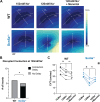
Scn5a heterozygous null (Scn5a+/-) mice have historically been used to investigate arrhythmogenic mechanisms of diseases such as Brugada syndrome (BrS) and Lev's disease. Previously, we demonstrated that reducing ephaptic coupling (EpC) in ex vivo hearts exacerbates pharmacological voltage-gated sodium channel (Nav)1.5 loss of function (LOF). Whether this effect is consistent in a genetic Nav1.5 LOF model is yet to be determined. We hypothesized that loss of EpC would result in greater reduction in conduction velocity (CV) for the Scn5a+/- mouse relative to wild type (WT). In vivo ECGs and ex vivo optical maps were recorded from Langendorff-perfused Scn5a+/- and WT mouse hearts. EpC was reduced with perfusion of a hyponatremic solution, the clinically relevant osmotic agent mannitol, or a combination of the two. Neither in vivo QRS duration nor ex vivo CV during normonatremia was significantly different between the two genotypes. In agreement with our hypothesis, we found that hyponatremia severely slowed CV and disrupted conduction for 4/5 Scn5a+/- mice, but 0/6 WT mice. In addition, treatment with mannitol slowed CV to a greater extent in Scn5a+/- relative to WT hearts. Unexpectedly, treatment with mannitol during hyponatremia did not further slow CV in either genotype, but resolved the disrupted conduction observed in Scn5a+/- hearts. Similar results in guinea pig hearts suggest the effects of mannitol and hyponatremia are not species specific. In conclusion, loss of EpC through either hyponatremia or mannitol alone results in slowed or disrupted conduction in a genetic model of Nav1.5 LOF. However, the combination of these interventions attenuates conduction slowing.NEW & NOTEWORTHY Cardiac sodium channel loss of function (LOF) diseases such as Brugada syndrome (BrS) are often concealed. We optically mapped mouse hearts with reduced sodium channel expression (Scn5a+/-) to evaluate whether reduced ephaptic coupling (EpC) can unmask conduction deficits. Data suggest that conduction deficits in the Scn5a+/- mouse may be unmasked by treatment with hyponatremia and perinexal widening via mannitol. These data support further investigation of hyponatremia and mannitol as novel diagnostics for sodium channel loss of function diseases.
Keywords: basic science research; congenital heart disease; electrophysiology; translational studies.
Conflict of interest statement
No conflicts of interest, financial or otherwise, are declared by the authors.
Figures


Similar articles
-
Arrhythmic substrate, slowed propagation and increased dispersion in conduction direction in the right ventricular outflow tract of murine Scn5a+/- hearts.Acta Physiol (Oxf). 2014 Aug;211(4):559-73. doi: 10.1111/apha.12324. Epub 2014 Jul 9. Acta Physiol (Oxf). 2014. PMID: 24913289 Free PMC article.
-
Normal interventricular differences in tissue architecture underlie right ventricular susceptibility to conduction abnormalities in a mouse model of Brugada syndrome.Cardiovasc Res. 2018 Apr 1;114(5):724-736. doi: 10.1093/cvr/cvx244. Cardiovasc Res. 2018. PMID: 29267949 Free PMC article.
-
Effects of flecainide and quinidine on arrhythmogenic properties of Scn5a+/- murine hearts modelling the Brugada syndrome.J Physiol. 2007 May 15;581(Pt 1):255-75. doi: 10.1113/jphysiol.2007.128785. Epub 2007 Feb 15. J Physiol. 2007. PMID: 17303635 Free PMC article.
-
Clinical Spectrum of SCN5A Mutations: Long QT Syndrome, Brugada Syndrome, and Cardiomyopathy.JACC Clin Electrophysiol. 2018 May;4(5):569-579. doi: 10.1016/j.jacep.2018.03.006. Epub 2018 May 2. JACC Clin Electrophysiol. 2018. PMID: 29798782 Review.
-
Sodium channel haploinsufficiency and structural change in ventricular arrhythmogenesis.Acta Physiol (Oxf). 2016 Feb;216(2):186-202. doi: 10.1111/apha.12577. Epub 2015 Sep 24. Acta Physiol (Oxf). 2016. PMID: 26284956 Review.
Cited by
-
Ultrastructure and cardiac impulse propagation: scaling up from microscopic to macroscopic conduction.J Physiol. 2025 Mar;603(7):1887-1901. doi: 10.1113/JP287632. Epub 2024 Nov 29. J Physiol. 2025. PMID: 39612369 Free PMC article. Review.
References
-
- Royer A, van Veen TAB, Le Bouter S, Marionneau C, Griol-Charhbili V, Léoni A-L, Steenman M, van Rijen HVM, Demolombe S, Goddard CA, Richer C, Escoubet B, Jarry-Guichard T, Colledge WH, Gros D, de Bakker JMT, Grace AA, Escande D, Charpentier F. Mouse model of SCN5A-linked hereditary Lenègre’s disease: age-related conduction slowing and myocardial fibrosis. Circulation 111: 1738–1746, 2005. doi:10.1161/01.CIR.0000160853.19867.61. - DOI - PubMed
-
- Jeevaratnam K, Zhang Y, Guzadhur L, Duehmke RM, Lei M, Grace AA, Huang CL-H. Differences in sino-atrial and atrio-ventricular function with age and sex attributable to the Scn5a+/− mutation in a murine cardiac model. Acta Physiol (Oxf) 200: 23–33, 2010. doi:10.1111/j.1748-1716.2010.02110.x. - DOI - PubMed
-
- van Veen TAB, Stein M, Royer A, Le Quang K, Charpentier F, Colledge WH, Huang CL-H, Wilders R, Grace AA, Escande D, de Bakker JMT, van Rijen HVM. Impaired impulse propagation in Scn5a-knockout mice: combined contribution of excitability, connexin expression, and tissue architecture in relation to aging. Circulation 112: 1927–1935, 2005. doi:10.1161/CIRCULATIONAHA.105.539072. - DOI - PubMed
-
- Jeevaratnam K, Poh Tee S, Zhang Y, Rewbury R, Guzadhur L, Duehmke R, Grace AA, Lei M, Huang CL-H. Delayed conduction and its implications in murine Scn5a+/− hearts: Independent and interacting effects of genotype, age, and sex. Pflugers Arch 461: 29–44, 2011. doi:10.1007/s00424-010-0906-1. - DOI - PMC - PubMed
Publication types
MeSH terms
Substances
Grants and funding
- R01HL102298/HHS | NIH | National Heart, Lung, and Blood Institute (NHLBI)
- R01 HL102298/HL/NHLBI NIH HHS/United States
- F31HL160172/HHS | NIH | National Heart, Lung, and Blood Institute (NHLBI)
- R01HL159097/HHS | NIH | National Heart, Lung, and Blood Institute (NHLBI)
- R01 HL159097/HL/NHLBI NIH HHS/United States
LinkOut - more resources
Full Text Sources
Molecular Biology Databases
Miscellaneous

