. 2024 Apr;13(10):e2303593.
doi: 10.1002/adhm.202303593. Epub 2024 Jan 22.

Near-Infrared Induced miR-34a Delivery from Nanoparticles in Esophageal Cancer Treatment

Affiliations

Near-Infrared Induced miR-34a Delivery from Nanoparticles in Esophageal Cancer Treatment

Nick A Alden et al. Adv Healthc Mater. 2024 Apr.

Abstract

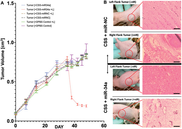
Current nucleic acid delivery methods have not achieved efficient, non-toxic delivery of miRNAs with tumor-specific selectivity. In this study, a new delivery system based on light-inducible gold-silver-gold, core-shell-shell (CSS) nanoparticles is presented. This system delivers small nucleic acid therapeutics with precise spatiotemporal control, demonstrating the potential for achieving tumor-specific selectivity and efficient delivery of miRNA mimics. The light-inducible particles leverage the photothermal heating of metal nanoparticles due to the local surface plasmonic resonance for controlled chemical cleavage and release of the miRNA mimic payload. The CSS morphology and composition result in a plasmonic resonance within the near-infrared (NIR) region of the light spectrum. Through this method, exogenous miR-34a-5p mimics are effectively delivered to human squamous cell carcinoma TE10 cells, leading to apoptosis induction without adverse effects on untransformed keratinocytes in vitro. The CSS nanoparticle delivery system is tested in vivo in Foxn1nu athymic nude mice with bilateral human esophageal TE10 cancer cells xenografts. These experiments reveal that this CSS nanoparticle conjugates, when systemically administered, followed by 850 nm light emitting diode irradiation at the tumor site, 6 h post-injection, produce a significant and sustained reduction in tumor volume, exceeding 87% in less than 72 h.

Keywords: Diels–Alder; esophageal cancer; miR‐34a; near‐infrared; plasmonic nanoparticles.


Conflict of interest statement

The authors declare no conflict of interest.

Figures

Figure 1
Figure 1
Targets of miR‐34a‐5p including ROCK1, TGIF2, MYC, and STAT3 in squamous cell cancer, and additional interacting pathways identifying the selection of miR‐34a as a potential therapeutic modulation of carcinogenic gene pathways in esophageal squamous cell carcinomas to drive discriminating apoptosis. Octagonal symbols indicate cascades reported to diminish normal cellular function. Solid and dotted arrows represent protein–protein (or related) interactions, and transcription/regulation (or related) interactions, respectively. Molecular symbols differentiate between the symbol's gene/protein/molecule class (p < 0.00005). Plus (+) symbols indicate positive interaction leading to oncogenesis; Negative (−) symbols indicate negative interactions leading to oncogenesis.
Figure 2
Figure 2
A) Nanoparticle size distribution from synthesis of the base, gold‐core seed, through shell formation as measured by DLS. B) Nanoparticle morphology and elemental composition detected using TEM equipped with EDS technology. Scale bar 50 nm. Within the STEM/EDS images, red color represents elemental gold (Au), and green color represents silver (Ag). C) Nanoparticle charge measured through zeta‐potential analysis. D) Nanoparticle mobility measured through zeta‐potential analysis. E) Colloidal stability of CSS‐miR34a nanoparticles in solution at varied times post‐synthesis at 20 °C (room temperature). n = 3 *Significant difference (p < 0.05). **Significant difference (p < 0.01). ***Significant difference (p < 0.001).
Figure 3
Figure 3
A) Schematic representation of the CSS nanoparticle, linker surface functionalization, and light activation of the reverse‐Diels–Alder reaction releasing siRNA payloads. B) Energy required for the activation of the reverse‐Diels–Alder reaction at the CSS nanoparticle surface due to plasmonic resonance at 850 nm absorbance. n = 3. *Significant difference (p < 0.05). ****Significant difference (p < 0.0001). C) Surface absorbance of the core, core–shell, and CSS nanoparticle through synthesis, where resonant wavelengths are denoted by the dotted line.
Figure 4
Figure 4
A) Fluorescent quantification of TE10 cells without PE‐Cy5 tagged CSS‐FDA‐miR34a nanoparticles through Flow Cytometry analysis. B) Fluorescent quantification of TE10 cells treated with PE‐Cy5 tagged CSS‐FDA‐miR34a nanoparticles through flow cytometry analysis. C) Fluorescent images of TE10 cells with CSS‐FDA‐miR34a mimic nanoparticles that received no irradiation at 850 nm, and imaged through confocal microscopy. Scale bar 10 µm. Blue, NucBlue/DAPI; Red, Cell‐Tracker Red, Green, FAM. D) Fluorescent images of TE10 cells treated with FAM‐tagged CSS‐FDA‐miR34a nanoparticles and irradiated with 850 nm for miRNA mimic release and imaged through confocal microscopy. Scale bar 10 µm.
Figure 5
Figure 5
A) Cell viability of TE10 cells treated with the CSS‐miRNA treatment groups measured using LIVE|DEAD fluorescent‐based assay for light‐based microscopy. The color green indicates the presence of living cells, while the color red indicates that the cells have died. B) Image quantification analysis of the fluorescent assay images shown in (A). Color‐scaled bars represent decreasing viability, bright green to dark green, respectively, and light pink to deep red respectively. C) Cell viability among TE10 cells treated with CSS‐miRNA mimic nanoparticle groups measured through fluorescent‐based, direct dsDNA quantification. “+L” indicates that NIR irradiation occurred for the corresponding treatment group. n = 3. *Significant difference (p < 0.05). ***Significant difference (p < 0.001). ****Significant difference (p < 0.0001).
Figure 6
Figure 6
Gene expression and target suppression analysis in vitro. A) Quantitative gene expression analysis of bioinformatics‐predicted miR34a targets measured through RT‐PCR to detect targets, ROCK1, TGIF2, MYC, and STAT3. n = 3. **Significant difference (p < 0.01). ****Significant difference (p < 0.0001). B) Qualitative analysis of intracellular levels of caspase‐3, known to direct apoptotic cell signaling pathways, using western blot to detect the presence of proCASP3 (inactive caspase‐3), and actCASP3 (active caspase‐3) in TE10 treatment samples.
Figure 7
Figure 7
A) Quantification of tumor volume over time before, during, and after treatment with the CSS‐miRNA nanoparticle treatment groups. n = 6. B) Histological analysis of mice treated with the CSS‐miRNA treatment groups. H&E‐stained sections imaged under light‐based microscopy (Scale bar is 2 mm). The extracellular matrices are stained pink, cell nuclei in purple, and empty space in white.

Similar articles

Cited by

References

    1. Trotti A., Byhardt R., Stetz J., Gwede C., Corn B., Fu K., Gunderson L., McCormick B., Morrisintegral M., Rich T., Shipley W., Curran W., Int J Radiat Oncol Biol Phys 2000, 47, 13. - PubMed
    1. Carelle N., Piotto E., Bellanger A., Germanaud J., Thuillier A., Khayat D., Cancer 2002, 95, 155. - PubMed
    1. Pattabiraman D. R., Weinberg R. A., Nat Rev Drug Discov 2014, 13, 497. - PMC - PubMed
    1. Cancer Facts and Figures 2022, American Cancer Society, 2022, https://www.cancer.org/research/cancer‐facts‐statistics/all‐cancer‐facts... (accessed: Febuary 2023).
    1. Wang Y., Grainger D. W., Front Chem Sci Eng 2014, 8, 265.

Publication types