

Reconstruction of lower limb defect with a variant sural neuro-fasciocutaneous flap: A case report
- PMID: 38215575
- PMCID: PMC10821621
- DOI: 10.1016/j.ijscr.2024.109236
Reconstruction of lower limb defect with a variant sural neuro-fasciocutaneous flap: A case report
Abstract
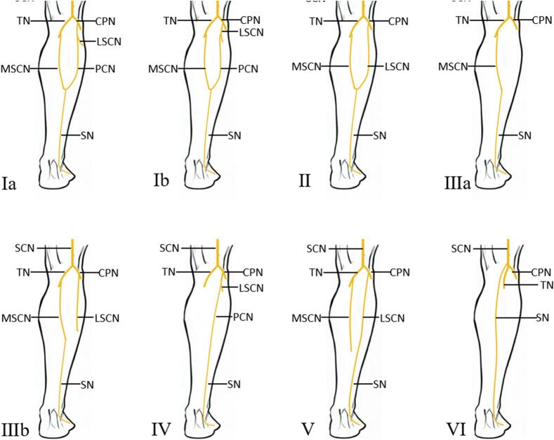
Introduction: The sural neuro-fasciocutaneous flap is widely used for reconstructing skin defects in the lower calf. Variations of the sural nerve in the calf are infrequent, which may require a variation in the traditional surgical procedure.
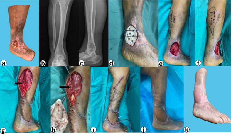
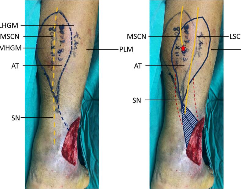
Case presentation: A 76-year-old male patient had soft tissue defect of the right lateral ankle and lower leg caused by an accident 18 years ago. He had exposed bones and had osteomyelitis. He underwent two primary operations, and finally, we used a sural neuro-fasciocutaneous flap to effectively cover the defect. We observed that the course of the sural nerve was atypical during the surgery, and we adjusted the flap axis laterally to bring the lateral sural cutaneous nerve inside the flap to improve the success rate of the surgery. The flap entirely survived, and there was no sensory impairment in the calf. The patient was discharged from the hospital after 10 days.
Clinical discussion: Some type of variant of the sural nerve makes the flap harvest without the neurovascular component of the sural nerve and the cutaneous chain, which might decrease flap survival. Moving the flap axis laterally and bringing in the lateral sural nerve or peroneal communicating nerve offers an adequate blood supply to the vascular territory and the flap region.
Conclusion: In patients with sural nerve variants, the procedure does not have to follow the traditional theory of the sural neuro-fasciocutaneous flap. Preoperative and intraoperative protection of the sural nerve variant should also be considered.
Keywords: Case report; Lateral calf defect; Reconstruction; Sural neuro-fasciocutaneous flap; Variant.
Copyright © 2024 The Authors. Published by Elsevier Ltd.. All rights reserved.
Conflict of interest statement
Declaration of competing interest The authors declare no conflict of interest.
Figures
Similar articles
-
Non-microsurgical bipedicled reverse sural fasciocutaneous flap with preservation of medial and lateral sural cutaneous nerve: Current surgical management of skin defect after traumatic Achilles tendon rupture - A case report.Int J Surg Case Rep. 2021 Jan;78:259-264. doi: 10.1016/j.ijscr.2020.12.027. Epub 2020 Dec 16. Int J Surg Case Rep. 2021. PMID: 33373919 Free PMC article.
-
The distally based lateral sural neuro-lesser saphenous veno-fasciocutaneous flap: anatomical basis and clinical applications.J Orthop Traumatol. 2014 Sep;15(3):215-23. doi: 10.1007/s10195-012-0202-2. Epub 2012 Jun 26. J Orthop Traumatol. 2014. PMID: 22733171 Free PMC article.
-
Distally based sural neuro-lesser saphenous veno-fasciocutaneous compound flap with a low rotation point: microdissection and clinical application.Ann Plast Surg. 2009 Apr;62(4):395-404. doi: 10.1097/SAP.0b013e31816dd3a9. Ann Plast Surg. 2009. PMID: 19325344
-
Modified distally based sural neuro-veno-fasciocutaneous flap: anatomical study and clinical applications.Microsurgery. 2005;25(7):543-50. doi: 10.1002/micr.20162. Microsurgery. 2005. PMID: 16178006
-
Morphological Variability of the Sural Nerve and Its Clinical Significance.J Clin Med. 2024 Oct 11;13(20):6055. doi: 10.3390/jcm13206055. J Clin Med. 2024. PMID: 39458004 Free PMC article. Review.
References
-
- Martinengo L., Olsson M., Bajpai R., Soljak M., Upton Z., Schmidtchen A., Car J., Järbrink K. Prevalence of chronic wounds in the general population: systematic review and meta-analysis of observational studies. Ann. Epidemiol. 2019;29:8–15. doi: 10.1016/j.annepidem.2018.10.005. January. - DOI - PubMed
-
- Vos T., Allen C., Arora M., Barber R.M., Bhutta Z.A., Brown A., Carter A., Casey D.C., Charlson F.J., Chen A.Z., Coggeshall M., Cornaby L., Dandona L., Dicker D.J., Dilegge T., Erskine H.E., Ferrari A.J., Fitzmaurice C., Fleming T., Murray C.J.L. Global, regional, and national incidence, prevalence, and years lived with disability for 310 diseases and injuries, 1990–2015: a systematic analysis for the Global Burden of Disease Study 2015. Lancet. 2016;388(10053):1545–1602. doi: 10.1016/s0140-6736(16)31678-6. October. - DOI - PMC - PubMed
-
- Gould L., Abadir P., Brem H., Carter M., Conner-Kerr T., Davidson J., DiPietro L., Falanga V., Fife C., Gardner S., Grice E., Harmon J., Hazzard W.R., High K.P., Houghton P., Jacobson N., Kirsner R.S., Kovacs E.J., Margolis D., Schmader K. Chronic wound repair and healing in older adults: current status and future research. J. Am. Geriatr. Soc. 2015;63(3):427–438. doi: 10.1111/jgs.13332. March. - DOI - PMC - PubMed
Publication types
LinkOut - more resources
Full Text Sources
