

Comparing the efficacy of awake and sedated MEG to TMS in mapping hand sensorimotor cortex in a clinical cohort
- PMID: 38215622
- PMCID: PMC10821581
- DOI: 10.1016/j.nicl.2024.103562
Comparing the efficacy of awake and sedated MEG to TMS in mapping hand sensorimotor cortex in a clinical cohort
Abstract
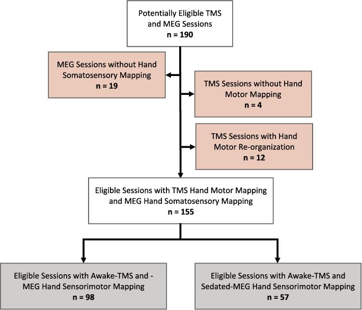
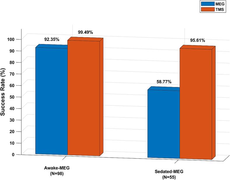
Non-invasive methods such as Transcranial Magnetic Stimulation (TMS) and magnetoencephalography (MEG) aid in the pre-surgical evaluation of patients with epilepsy or brain tumor to identify sensorimotor cortices. MEG requires sedation in children or patients with developmental delay. However, TMS can be applied to awake patients of all ages with any cognitive abilities. In this study, we compared the efficacy of TMS with MEG (in awake and sedated states) in identifying the hand sensorimotor areas in patients with epilepsy or brain tumors. We identified 153 patients who underwent awake- (n = 98) or sedated-MEG (n = 55), along with awake TMS for hand sensorimotor mapping as part of their pre-surgical evaluation. TMS involved stimulating the precentral gyrus and recording electromyography responses, while MEG identified the somatosensory cortex during median nerve stimulation. Awake-MEG had a success rate of 92.35 % and TMS had 99.49 % (p-value = 0.5517). However, in the sedated-MEG cohort, TMS success rate of 95.61 % was significantly higher compared to MEG's 58.77 % (p-value = 0.0001). Factors affecting mapping success were analyzed. Logistic regression across the entire cohort identified patient sedation as the lone significant predictor, contrary to age, lesion, metal, and number of antiseizure medications (ASMs). A subsequent analysis replaced sedation with anesthetic drug dosage, revealing no significant predictors impacting somatosensory mapping success under sedation. This study yields insights into the utility of TMS and MEG in mapping hand sensorimotor cortices and underscores the importance of considering factors that influence eloquent cortex mapping limitations during sedation.
Keywords: Brain tumor; Epilepsy; Magnetoencephalography (MEG); Sedation; Sensorimotor mapping; Transcranial Magnetic Stimulation (TMS).
Copyright © 2024. Published by Elsevier Inc.
Conflict of interest statement
Declaration of competing interest The authors declare that they have no known competing financial interests or personal relationships that could have appeared to influence the work reported in this paper.
Figures
Similar articles
-
Assessing motor function in young children with transcranial magnetic stimulation.Pediatr Neurol. 2015 Jan;52(1):94-103. doi: 10.1016/j.pediatrneurol.2014.08.031. Epub 2014 Sep 18. Pediatr Neurol. 2015. PMID: 25439485
-
Localization of Sensorimotor Cortex Using Navigated Transcranial Magnetic Stimulation and Magnetoencephalography.Brain Topogr. 2019 Sep;32(5):873-881. doi: 10.1007/s10548-019-00716-w. Epub 2019 May 15. Brain Topogr. 2019. PMID: 31093863 Free PMC article.
-
Preoperative multimodal motor mapping: a comparison of magnetoencephalography imaging, navigated transcranial magnetic stimulation, and direct cortical stimulation.J Neurosurg. 2012 Aug;117(2):354-62. doi: 10.3171/2012.5.JNS112124. Epub 2012 Jun 15. J Neurosurg. 2012. PMID: 22702484 Free PMC article.
-
Neuromagnetic integrated methods tracking human brain mechanisms of sensorimotor areas 'plastic' reorganisation.Brain Res Brain Res Rev. 2000 Sep;33(2-3):131-54. doi: 10.1016/s0169-328x(00)00090-5. Brain Res Brain Res Rev. 2000. PMID: 11011062 Review.
-
On the relative merits of invasive and non-invasive pre-surgical brain mapping: New tools in ablative epilepsy surgery.Epilepsy Res. 2018 May;142:153-155. doi: 10.1016/j.eplepsyres.2017.07.002. Epub 2017 Jul 3. Epilepsy Res. 2018. PMID: 28716297 Review.
References
-
- Anderson C.T., Carlson C.E., Li Z., Raghavan M. Magnetoencephalography in the preoperative evaluation for epilepsy surgery. Curr. Neurol. Neurosci. Rep. 2014;14:1–8. - PubMed
-
- Balakrishnan G., Grover K.M., Mason K., Smith B., Barkley G.L., Tepley N., Bowyer S.M. A retrospective analysis of the effect of general anesthetics on the successful detection of interictal epileptiform activity in magnetoencephalography. Anesth. Analg. 2007;104:1493–1497. - PubMed
-
- Bercovici E., Pang E.W., Sharma R., Mohamed I.S., Imai K., Fujimoto A., Ochi A., Viljoen A., Chu B., Holowka S. Somatosensory-evoked fields on magnetoencephalography for epilepsy infants younger than 4 years with total intravenous anesthesia. Clin. Neurophysiol. 2008;119:1328–1334. - PubMed
-
- Birg L., Narayana S., Rezaie R., Papanicolaou A. Technical tips: MEG and EEG with sedation. Neurodiagnostic J. 2013;53:229–240. - PubMed
-
- Bowyer S.M., Pang E.W., Huang M., Papanicolaou A.C., Lee R.R. Presurgical functional mapping with magnetoencephalography. Neuroimaging Clin. 2020;30:159–174. - PubMed
