Magnetic resonance imaging improves diagnosis of placenta accreta spectrum requiring hysterectomy compared to ultrasound
- PMID: 38216054
- PMCID: PMC11070455
- DOI: 10.1016/j.ajogmf.2024.101280
Magnetic resonance imaging improves diagnosis of placenta accreta spectrum requiring hysterectomy compared to ultrasound
Abstract
Background: Magnetic resonance imaging has been used increasingly as an adjunct for ultrasound imaging for placenta accreta spectrum assessment and preoperative surgical planning, but its value has not been established yet. The ultrasound-based placenta accreta index is a well-validated standardized approach for placenta accreta spectrum evaluation. Placenta accreta spectrum-magnetic resonance imaging markers have been outlined in a joint guideline from the Society of Abdominal Radiology and the European Society of Urogenital Radiology.
Objective: This study aimed to compare placenta accreta spectrum-magnetic resonance imaging parameters with the ultrasound-based placenta accreta index in pregnancies at high risk for placenta accreta spectrum and to assess the additional diagnostic value of magnetic resonance imaging for placenta accreta spectrum that requires a cesarean hysterectomy.
Study design: This was a single-center, retrospective study of pregnant patients who underwent magnetic resonance imaging, in addition to ultrasonography, because of suspected placenta accreta spectrum. The ultrasound-based placenta accreta index and placenta accreta spectrum-magnetic resonance imaging parameters were obtained. Student's t test and Fisher's exact test were used to compare the groups in terms of the primary outcome (hysterectomy vs no hysterectomy). The diagnostic performance of magnetic resonance imaging and the ultrasound-based placenta accreta index was assessed using multivariable logistic regressions, receiver operating characteristics curves, the DeLong test, McNemar test, and the relative predictive value test.
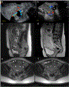
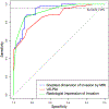
Results: A total of 82 patients were included in the study, 41 of whom required a hysterectomy. All patients who underwent a hysterectomy met the International Federation of Gynecology and Obstetrics clinical evidence of placenta accreta spectrum at the time of delivery. Multiple parameters of the ultrasound-based placenta accreta index and placenta accreta spectrum-magnetic resonance imaging were able to predict hysterectomy, and the parameter of greatest dimension of invasion by magnetic resonance imaging was the best quantitative predictor. At 96% sensitivity for hysterectomy, the cutoff values were 3.5 for the ultrasound-based placenta accreta index and 2.5 cm for the greatest dimension of invasion by magnetic resonance imaging. Using this sensitivity, the parameter of greatest dimension of invasion measured by magnetic resonance imaging had higher specificity (P=.0016) and a higher positive predictive value (P=.0018) than the ultrasound-based placenta accreta index, indicating an improved diagnostic threshold.
Conclusion: In a suspected high-risk group for placenta accreta spectrum, magnetic resonance imaging identified more patients who will not need a hysterectomy than when using the ultrasound-based placenta accrete index only. Magnetic resonance imaging has the potential to aid patient counseling, surgical planning, and delivery timing, including preterm delivery decisions for patients with placenta accreta spectrum requiring hysterectomy.
Keywords: MRI; hysterectomy; placenta accreta index; placenta accreta spectrum; ultrasound.
Copyright © 2024 Elsevier Inc. All rights reserved.
Conflict of interest statement
The authors report no conflict of interest.
Figures


References
-
- American College of Obstetricians and Gynecologists, Society for Maternal-Fetal Medicine. Obstetric care consensus no. 7: placenta accreta spectrum. Obstet Gynecol 2018;132: e259–75. - PubMed
-
- Sentilhes L, Kayem G, Chandraharan E, Palacios-Jaraquemada J, Jauniaux E. FIGO Placenta Accreta Diagnosis and Management Expert Consensus Panel. FIGO consensus guidelines on placenta accreta spectrum disorders: conservative management. Int J Gynaecol Obstet 2018;140:291–8. - PubMed
-
- Fleming ET, Yule CS, Lafferty AK, Happe SK, McIntire DD, Spong CY. Changing patterns of peripartum hysterectomy over time. Obstet Gynecol 2021;138:799–801. - PubMed
-
- Shamshirsaz AA, Fox KA, Erfani H, et al. Outcomes of planned compared with urgent deliveries using a multidisciplinary team approach for morbidly adherent placenta. Obstet Gynecol 2018;131:234–41. - PubMed
-
- Shamshirsaz AA, Fox KA, Salmanian B, et al. Maternal morbidity in patients with morbidly adherent placenta treated with and without a standardized multidisciplinary approach. Am J Obstet Gynecol 2015;212:218.e1–9. - PubMed
Publication types
MeSH terms
Grants and funding
LinkOut - more resources
Full Text Sources
Miscellaneous

