. 2024 Feb 7;45(2):224-228.
doi: 10.3174/ajnr.A8105.

White Matter Injury on Early-versus-Term-Equivalent Age Brain MRI in Infants Born Preterm

Affiliations

White Matter Injury on Early-versus-Term-Equivalent Age Brain MRI in Infants Born Preterm

Sriya Roychaudhuri et al. AJNR Am J Neuroradiol. .

Abstract

Background and purpose: White matter injury in infants born preterm is associated with adverse neurodevelopmental outcomes, depending on the extent and location. White matter injury can be visualized with MR imaging in the initial weeks following preterm birth but is more commonly defined at term-equivalent-age MR imaging. Our aim was to see how white matter injury detection in MR imaging compares between the 2 time points.

Materials and methods: This study compared white matter injury on early brain MR imaging (30-34 weeks' postmenstrual age) with white matter injury assessment at term-equivalent (37-42 weeks) MR imaging, using 2 previously published and standardized scoring systems, in a cohort of 30 preterm infants born at <33 weeks' gestational age.

Results: There was a strong association between the systematic assessments of white matter injury at the 2 time points (P = .007) and the global injury severity (P < .001).

Conclusions: Although the optimal timing to undertake neuroimaging in the preterm infant remains to be determined, both early (30-34 weeks) and term-equivalent MR imaging provide valuable information on white matter injury and the risk of associated sequelae.


Figures

FIG 1.
FIG 1.
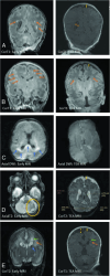
WMI as appreciated in early (1T scanner) versus TEA MR imaging (3T scanner) in different cases. A, The image on the left shows an early MR imaging and the extent of WMI (orange arrows) and its evolution at TEA (image on the right) in which there is an absence of visible WM lesions. WMI sequelae can be appreciated as volume loss, with arrows indicating a dilated ventricle and enlarged subarachnoid space (yellow arrow). B, Early T1 images show severe WMI (orange arrows) with reduced conspicuity at TEA but with some volume loss. C, Early MR imaging at day 3 of life shows diffusion restriction in WM (blue arrows), which has resolved at TEA. D, Isolated unilateral cerebellar hemorrhage (encircled in yellow) with eventual disparity in cerebellar hemispheric sizes and bilateral cerebral WM volume loss at TEA. E, Early IVH (blue arrow) with dilation resulting in periventricular WMI (orange arrows) with cyst formation (green arrows) and volume loss (yellow arrows) at TEA.
FIG 2.
FIG 2.
Evolution of WMI severity categories from early to TEA MRI. The thickness of arrows is proportional to the number of cases.

Comment in

  • On Behalf of Serial Imaging in Preterm Infants.
    Saraiva J, Soares-Fernandes JP. Saraiva J, et al. AJNR Am J Neuroradiol. 2024 Jun 7;45(6):E16. doi: 10.3174/ajnr.A8223. AJNR Am J Neuroradiol. 2024. PMID: 38782591 Free PMC article. No abstract available.

References

    1. Rees P, Callan C, Chadda KR, et al. . Preterm brain injury and neurodevelopmental outcomes: a meta-analysis. Pediatrics 2022;150:e2022057442 10.1542/peds.2022-057442 - DOI - PMC - PubMed
    1. Novak I, Morgan C, Adde L, et al. . Early, accurate diagnosis and early intervention in cerebral palsy: advances in diagnosis and treatment. JAMA Pediatr 2017;171:897–907 10.1001/jamapediatrics.2017.1689 - DOI - PMC - PubMed
    1. Inder TE, Volpe JJ, Anderson PJ. Defining the neurologic consequences of preterm birth. N Engl J Med 2023;389:441–53 10.1056/NEJMra2303347 - DOI - PubMed
    1. de Bruïne FT, van Wezel-Meijler G. Radiological assessment of white matter injury in very preterm infants. Imaging Med 2012;4:541–50 10.2217/iim.12.37 - DOI
    1. Kidokoro H, Neil JJ, Inder TE. New MR imaging assessment tool to define brain abnormalities in very preterm infants at term. AJNR Am J Neuroradiol 2013;34:2208–14 10.3174/ajnr.A3521 - DOI - PMC - PubMed

LinkOut - more resources