. 2024 Jan 12;15(1):477.
doi: 10.1038/s41467-024-44755-9.

Functional interactions between neurofibromatosis tumor suppressors underlie Schwann cell tumor de-differentiation and treatment resistance

Affiliations

Functional interactions between neurofibromatosis tumor suppressors underlie Schwann cell tumor de-differentiation and treatment resistance

Harish N Vasudevan et al. Nat Commun. .

Abstract

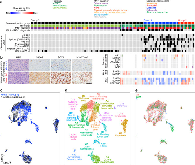
Schwann cell tumors are the most common cancers of the peripheral nervous system and can arise in patients with neurofibromatosis type-1 (NF-1) or neurofibromatosis type-2 (NF-2). Functional interactions between NF1 and NF2 and broader mechanisms underlying malignant transformation of the Schwann lineage are unclear. Here we integrate bulk and single-cell genomics, biochemistry, and pharmacology across human samples, cell lines, and mouse allografts to identify cellular de-differentiation mechanisms driving malignant transformation and treatment resistance. We find DNA methylation groups of Schwann cell tumors can be distinguished by differentiation programs that correlate with response to the MEK inhibitor selumetinib. Functional genomic screening in NF1-mutant tumor cells reveals NF2 loss and PAK activation underlie selumetinib resistance, and we find that concurrent MEK and PAK inhibition is effective in vivo. These data support a de-differentiation paradigm underlying malignant transformation and treatment resistance of Schwann cell tumors and elucidate a functional link between NF1 and NF2.


Conflict of interest statement

The authors declare no competing interests.

Figures

Fig. 1
Fig. 1. Multiplatform bulk and single-cell molecular profiling reveals de-differentiation underlies malignant transformation of human Schwann cell tumors.
a DNA methylation profiling and consensus k-means clustering using Spearman’s correlation of human schwannomas (n = 67), neurofibromas (n = 10), and malignant peripheral nerve sheath tumors (MPNSTs) (n = 42) reveals 3 Schwann cell tumor groups. Whole exome sequencing (n = 34), RNA sequencing (RNA-seq, n = 18), or immunohistochemistry (IHC, n = 36) of histological neurofibromas and MPNSTs shows distinct mutational patterns underlying epigenetic dysregulation and loss of Schwann cell differentiation markers (S100B, SOX10). MNP, molecular neuropathology classification. b Representative IHC images showing loss of Schwann cell differentiation markers or loss H3K27me in Group 1 compared to Group 2/3 histological neurofibromas and MPNSTs and repeated independently on all 36 tumor samples analyzed (scale bar, 100 μm). c Harmonized single-nuclear RNA sequencing uniform manifold approximation and projection (UMAP) of 19,276 nuclei annotated by tumor of origin from Group 1 MPNSTs (blue, n = 3) or Group 3 neurofibromas (black, n = 3). d Tumor and non-tumor cell types from single-nuclear RNA sequencing of Schwann cell tumors defined using a combination of automated cell type classification, cell signature gene sets from MSigDB, cluster marker genes, and cell cycle phase estimation. e Single-nuclear RNA sequencing cell cycle phase estimation demonstrating Group 1 MPNSTs are enriched in actively dividing cells (green, blue) while Group 3 neurofibromas are enriched for non-dividing cells (pink).
Fig. 2
Fig. 2. Schwann cell differentiation underlies MEK inhibitor response.
a RNA sequencing and hierarchical clustering using consensus PRC2 target genes distinguishes patient-derived neurofibroma or MPNST cells by PRC2-intact (red) or PRC2-mutant (blue) status with loss of Schwann cell differentiation markers (S100B, SOX10) in PRC2-mutant cells. b CRISPRi suppression of the PRC2 components SUZ12 (sgSUZ12) or EED (sgEED) using 2 separate sgRNAs each inhibits Schwann cell differentiation marker expression and leads to selumetinib resistance in NF1-mutant NF95.11b neurofibroma cells compared to non-targeted sgRNAs (sgNTC). SUZ12, EED, and S100B expression were analyzed using QPCR. Cell viability after 48 h of 1 μM selumetinib treatment was assessed using MTT assays and normalized to vehicle control treatments for each cell line (dotted line) (n = 3 biologically independent experiments for all conditions). c NF95.11b neurofibroma cell immunoblots reveal 1 μM selumetinib treatment transiently inhibits pERK and induces pMEK and apoptosis over time (n = 2 biologically independent experiments). d Single-cell RNA sequencing UMAP analysis of 26,608 cells from JW23.3 male MPNST allografts in NU/NU female recipient mice treated with 25 mg/kg selumetinib twice daily by oral gavage (n = 3) or vehicle control (n = 2) for 21 days. Non-tumor cells were filtered using Xist expression to identify female host cells. Tumor cells were defined using automated cell type classification, cell signature gene sets, cell cycle phase estimation, and cluster marker genes. e C0 (selumetinib resistant cells) and C1 (proliferating tumor cells) were enriched in selumetinib (n = 3 biologically independent mice) or vehicle (n = 2 biologically independent mice) treated allograft single-cell RNA sequencing samples, respectively. f Nf2 expression was significantly decreased in selumetinib compared to vehicle treated allograft single-cell RNA sequencing samples (p = 2.2 × 10−16, Wilcoxon rank sum test). Lines represent means. Error bars represent standard error of the means. *p < 0.05, **p < 0.01, ***p ≤ 0.0001, two sided Student’s t tests.
Fig. 3
Fig. 3. NF2 inactivation drives de-differentiation and MEK inhibitor resistance in NF1-mutant Schwann cell tumors.
a Volcano plot depicting significantly enriched sgRNAs (n = 563, red) or depleted sgRNAs (n = 608, blue) from triplicate genome-wide CRISPRi screens in NF1-mutant NF95.11b neurofibroma cells stably expressing dCas9-KRAB and treated with 1 μM selumetinib for 10 days compared to baseline transduction pre-treatment at T0. Significant hits mediating selumetinib resistance (red) or selumetinib sensitivity (blue) are shown. X-axis is normalized log2 sgRNA abundance count. b CRISPRi suppression of NF2 using 2 independent sgRNAs (gray, sgNF2) inhibits Schwann cell differentiation and drives selumetinib resistance in NF1-mutant NF95.11b neurofibroma cells stably expressing dCas9-KRAB compared to sgRNA non-targeting controls (sgNTC). NF2 or S100B expression were analyzed using RNA sequencing (TPM, transcripts per million) (n = 2 biologically independent experiments per sgRNA). Cell viability after 48 h of 1 μM selumetinib treatment was assessed using MTT assays and normalized to vehicle control treatments for each cell line (dotted line) (n = 4 biologically independent experiments). c NF95.11b neurofibroma cell immunoblots reveal sgRNAs suppressing NF2 induce PAK1 phosphorylation without altering pERK, pMEK, or pAKT compared to sgNTCs (n = 2 biologically independent experiments). d NF95.11b neurofibroma cell immunoblots from cells treated with combination molecular therapy inhibiting MEK (selumetinib) and PAK1 (NVS-PAK1-1) reveals robust biochemical repression of pERK in NF1-mutant, NF2-mutant cells. e Treatment of either JW23.3 or JW18.2 MPNST allografts in NU/NU mice with 25 mg/kg selumetinib twice daily by oral gavage (n = 6 biologically independent JW23.3 allografts, n = 10 biologically independent JW18.2 allografts) or 10 mg/kg NVS-PAK1-1 once daily by oral gavage (n = 4 biologically independent JW23.3 allografts, n = 10 biologically independent JW18.2 allografts) or combined selumetinib and NVS-PAK1-1 (n = 5 biologically independent JW23.3 allografts, n = 10 biologically independent JW18.2 allografts), or vehicle control (n = 5 JW23.3 allografts, n = 10 biologically independent JW18.2 allografts) for 21 days demonstrates combination molecular therapy blocks MPNST allograft growth compared to vehicle control or molecular monotherapy. f Schematic model summarizing genetic, biologic, and therapeutic mechanisms underlying Schwann cell tumor transformation. Lines represent means. Error bars represent standard error of the means. *p < 0.05, **p < 0.01, ***p ≤ 0.0001, two sided Student’s t tests.

References

    1. Ostrom QT, et al. CBTRUS statistical report: primary brain and other central nervous system tumors diagnosed in the United States in 2015–2019. Neuro Oncol. 2022;24:v1–v95. doi: 10.1093/neuonc/noac202. - DOI - PMC - PubMed
    1. Farid M, et al. Malignant peripheral nerve sheath tumors (MPNST) Oncologist. 2014;19:193–201. doi: 10.1634/theoncologist.2013-0328. - DOI - PMC - PubMed
    1. Vasudevan HN, Lucas CHG, Villanueva-Meyer JE, Theodosopoulos PV, Raleigh DR. Genetic events and signaling mechanisms underlying Schwann cell fate in development and cancer. Neurosurgery. 2021;88:234–245. doi: 10.1093/neuros/nyaa455. - DOI - PMC - PubMed
    1. Choudhury A, et al. Meningioma DNA methylation groups identify biological drivers and therapeutic vulnerabilities. Nat. Genet. 2022;54:649–659. doi: 10.1038/s41588-022-01061-8. - DOI - PMC - PubMed
    1. Gutmann DH, et al. Neurofibromatosis type 1. Nat. Rev. Dis. Prim. 2017;3:1–17. - PubMed

MeSH terms

Substances