. 2024 Jan 12;14(1):1199.
doi: 10.1038/s41598-023-50864-0.

Novel rapid method for identifying and quantifying pathogenic bacteria within four hours of blood collection

Affiliations

Novel rapid method for identifying and quantifying pathogenic bacteria within four hours of blood collection

Akio Miyakoshi et al. Sci Rep. .

Abstract

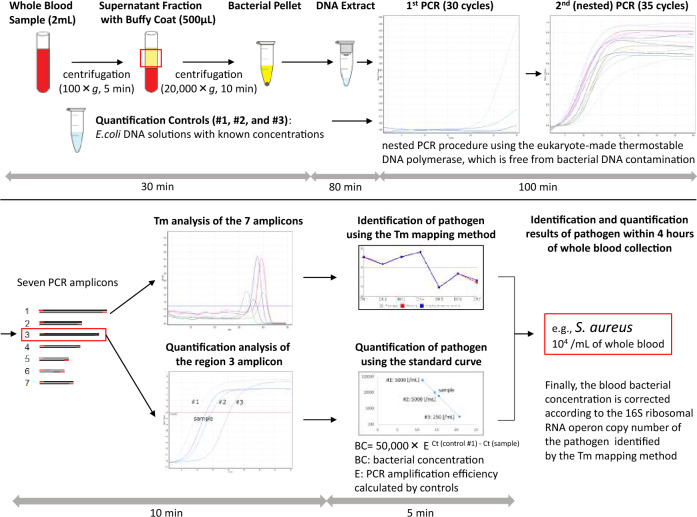
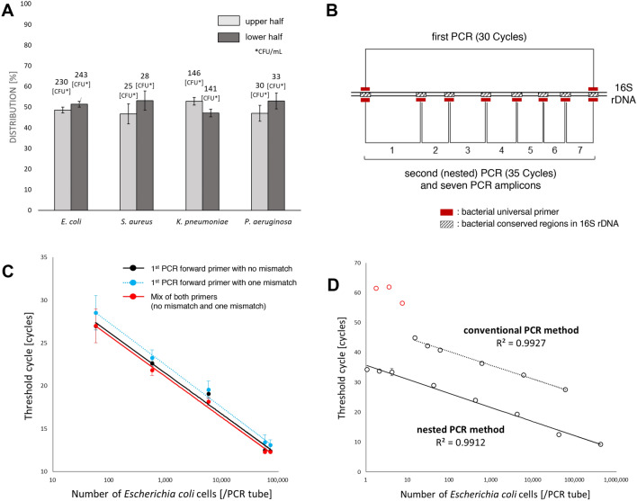
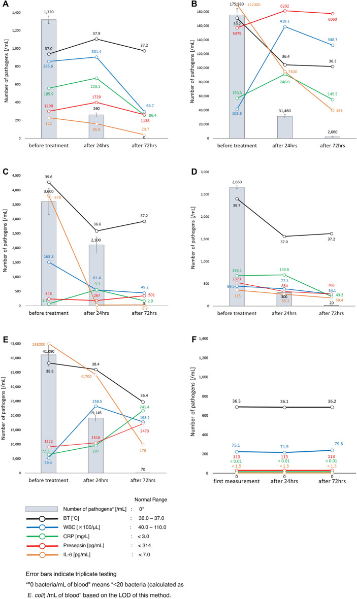
Sepsis is life-threatening organ dysfunction and is considered a major cause of health loss. However, since the current biomarkers of sepsis reflect the host's immune response to microorganisms, they would inevitably cause a time-lag. This means that there is still no truly reliable biomarker of sepsis. In the present study, we developed a novel method for identifying and quantifying unknown pathogenic bacteria within four hours of sample collection. The most important point of this study is that the novel method can be used to determine the number of bacteria in a sample as a novel biomarker of infectious diseases. Indeed, based on the number of bacteria, we were able to accurately estimate the severity of microbial infection. Furthermore, using the time-dependent changes in the number of bacteria, we were able to monitor the therapeutic effect accurately. The rapid identification and quantification of bacteria may change our approach to medical care.


Conflict of interest statement

The authors declare no competing interests.

Figures

Figure 1
Figure 1
Workflow of the novel rapid method for identifying and quantifying unknown pathogenic bacteria in sepsis within four hours of whole-blood collection. *This figure conceptually shows the workflow of the novel method, and the individual values in the figure have no meaning.
Figure 2
Figure 2
Establishment of the quantification method. (A) Distribution of bacteria (CFU/mL) in saline after low-speed centrifugation. The bacterial concentration in the upper half and the lower half were examined after low-speed centrifugation (100×g, 5 min). A total of 2 mL of bacterial suspension (E. coli, S. aureus, K. pneumoniae and P. aeruginosa) in saline was centrifuged, and the colony-forming unit (CFU) value was measured in the upper and lower half (1 mL each). The figure shows the ratio (%) of each half. Error bars indicate triplicate testing. (B) The primer designs. Nested PCR is performed using seven bacterial universal primer sets, and then the seven PCR amplicons (Tm values) are obtained. The pathogenic bacteria are identified using the seven Tm values of region 1 to 7 amplicons, and the bacterial concentration is measured using the region 3 amplicon alone. (C) Standard curves generated using three different primers. We generated standard curves by conducting an analysis of serial dilutions of E. coli DNA using primers as follows: 1st PCR forward primer with no mismatch against E. coli, 1st PCR forward primer with one mismatch against E. coli, and a mix of both 1st PCR forward primers (no mismatch : one mismatch = 1: 1). The 1st PCR reverse primer was the same in all cases. These results indicated that the quantification result using the 1st PCR forward primer with 1 mismatch was around 25% lower than that using the 1st PCR forward primer with no mismatch. In contrast, the quantification result using the mixed 1st PCR forward primers was almost the same as that using the 1st PCR forward primer with no mismatch. Error bars indicate triplicate testing. (D) The comparison of the regression lines generated by serial dilution of a known amount of E. coli DNA using conventional PCR method and nested PCR method. The regression lines calculated for the datum points are shown. The linear correlation between the Ct values and the logarithm of the number of E. coli/PCR tube (R2 value) was > 0.99. Error bars indicate triplicate testing.
Figure 3
Figure 3
Time-dependent changes of the number of bacteria, BT, WBC, CRP, Presepsin and IL-6 in sepsis patients before antibiotic treatment and 24 and 72 h after. (A) Case 1: A 76-year-old woman was diagnosed with sepsis associated with urinary tract infection. The Tm mapping result (Difference Value = 0.29) was E. coli, and 2 days later, the blood culture result and urine culture result were also consistent with E. coli. Meropenem (E. coli isolates were susceptible to meropenem) was then administered systemically. Error bars indicate triplicate testing. (B) Case 2: An 88-year-old woman was diagnosed with sepsis associated with retrograde cholangitis. The Tm mapping result (Difference Value > 0.5) was polymicrobial infection, and 2 days later, the blood culture result was found to be Klebsiella oxytoca, Haemophilus influenzae, and S. pneumoniae. The blood bacterial concentration was thus calculated in conversion as E. coli. Cefepime (K. oxytoca and H. influenzae isolates were susceptible to cefepime. S. pneumoniae isolates were intermediate to cefepime) was then administered systemically. Error bars indicate triplicate testing. (C) Case 3: A 94-year-old woman was diagnosed with sepsis associated with urinary tract infection. The Tm mapping result (Difference Value = 0.19) was E. coli, and 2 days later, the blood culture result and the urine culture result were also consistent with E. coli. Tazobactam/piperacillin (E. coli isolates were susceptible to tazobactam/piperacillin) was then administered systemically. Error bars indicate triplicate testing. (D) Case 4: An 84-year-old woman was diagnosed with sepsis associated with postoperative wound infection after the spine surgery. The Tm mapping result (Difference Value = 0.28) was S. dysgalactiae, and 2 days later, the blood culture result was also consistent with S. dysgalactiae. Tazobactam/piperacillin (S. dysgalactiae isolates were susceptible to tazobactam/piperacillin) was then administered systemically. Error bars indicate triplicate testing. (E) Case 5: An 81-year-old woman was diagnosed with sepsis associated with urinary tract infection. The Tm mapping result (Difference Value = 0.48) was Enterobacter aerogenes, and 2 days later, the blood culture result and the urine culture result were also consistent with E. aerogenes (2 types of mutant strains). Tazobactam/piperacillin (E. aerogenes isolates were susceptible to tazobactam/piperacillin) was then administered systemically. Error bars indicate triplicate testing. (F) Healthy Control: A 44-year-old woman who does not have any illnesses including infectious diseases and participated in this study as a volunteer. No bacteria were detected in the blood using the Tm mapping method and blood culture. Error bars indicate triplicate testing.

References

    1. Singer M, et al. The third international consensus definitions for sepsis and septic shock (Sepsis-3) JAMA. 2016;315:801–810. doi: 10.1001/jama.2016.0287. - DOI - PMC - PubMed
    1. Rudd KE, et al. Global, regional, and national sepsis incidence and mortality, 1990–2017: Analysis for the Global Burden of Disease Study. Lancet. 2020;395:200–211. doi: 10.1016/s0140-6736(19)32989-7. - DOI - PMC - PubMed
    1. Asner SA, Desgranges F, Schrijver IT, Calandra T. Impact of the timeliness of antibiotic therapy on the outcome of patients with sepsis and septic shock. J. Infect. 2021;82:125–134. doi: 10.1016/j.jinf.2021.03.003. - DOI - PubMed
    1. MacArthur RD, et al. Adequacy of early empiric antibiotic treatment and survival in severe sepsis: Experience from the MONARCS trial. Clin. Infect. Dis. 2004;38:284–288. doi: 10.1086/379825. - DOI - PubMed
    1. Niimi H, et al. Melting temperature mapping method: A novel method for rapid identification of unknown pathogenic microorganisms within three hours of sample collection. Sci. Rep. 2015;5:12543. doi: 10.1038/srep12543. - DOI - PMC - PubMed