

Lysine demethylase 5C inhibits transcription of prefoldin subunit 5 to activate c-Myc signal transduction and colorectal cancer progression
- PMID: 38216914
- PMCID: PMC10785505
- DOI: 10.1186/s10020-023-00775-7
Lysine demethylase 5C inhibits transcription of prefoldin subunit 5 to activate c-Myc signal transduction and colorectal cancer progression
Abstract
Background: Lysine demethylase 5C (KDM5C) has been implicated in the development of several human cancers. This study aims to investigate the role of KDM5C in the progression of colorectal cancer (CRC) and explore the associated molecular mechanism.
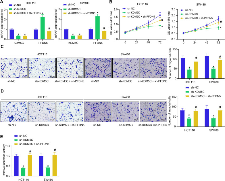
Methods: Bioinformatics tools were employed to predict the target genes of KDM5C in CRC. The expression levels of KDM5C and prefoldin subunit 5 (PFDN5) in CRC cells were determined by RT-qPCR and western blot assays. The interaction between KDM5C, H3K4me3, and PFDN5 was validated by chromatin immunoprecipitation. Expression and prognostic values of KDM5C and PFDN5 in CRC were analyzed in a cohort of 72 patients. The function of KDM5C/PFDN5 in c-Myc signal transduction was analyzed by luciferase assay. Silencing of KDM5C and PFDN5 was induced in CRC cell lines to analyze the cell malignant phenotype in vitro and tumorigenic activity in nude mice.
Results: KDM5C exhibited high expression, while PFDN5 displayed low expression in CRC cells and clinical CRC samples. High KDM5C levels correlated with poor survival and unfavorable clinical presentation, whereas elevated PFDN5 correlated with improved patient outcomes. KDM5C mediated demethylation of H3K4me3 on the PFDN5 promoter, suppressing its transcription and thereby enhancing the transcriptional activity of c-Myc. KDM5C knockdown in CRC cells suppressed cell proliferation, migration and invasion, epithelial-mesenchymal transition, and tumorigenic activity while increasing autophagy and apoptosis rates. However, the malignant behavior of cells was restored by the further silencing of PFDN5.
Conclusion: This study demonstrates that KDM5C inhibits PFDN5 transcription, thereby activating c-Myc signal transduction and promoting CRC progression.
Keywords: Apoptosis; Autophagy; Colorectal cancer; Lysine demethylase 5C; Prefoldin subunit 5.
© 2024. The Author(s).
Conflict of interest statement
The authors report no competing interest.
Figures


Similar articles
-
METTL14-mediated N6-methyladenosine modification of SOX4 mRNA inhibits tumor metastasis in colorectal cancer.Mol Cancer. 2020 Jun 17;19(1):106. doi: 10.1186/s12943-020-01220-7. Mol Cancer. 2020. PMID: 32552762 Free PMC article.
-
Histone Demethylase JMJD2D Interacts With β-Catenin to Induce Transcription and Activate Colorectal Cancer Cell Proliferation and Tumor Growth in Mice.Gastroenterology. 2019 Mar;156(4):1112-1126. doi: 10.1053/j.gastro.2018.11.036. Epub 2018 Nov 23. Gastroenterology. 2019. PMID: 30472235
-
HOXD3 Up-regulating KDM5C Promotes Malignant Progression of Diffuse Large B-Cell Lymphoma by Decreasing p53 Expression.Balkan Med J. 2022 Jan 25;39(1):30-38. doi: 10.5152/balkanmedj.2021.21068. Epub 2021 Dec 20. Balkan Med J. 2022. PMID: 34928233 Free PMC article.
-
Lysine demethylase 5C epigenetically reduces transcription of ITIH1 that results in augmented progression of liver hepatocellular carcinoma.Kaohsiung J Med Sci. 2022 May;38(5):437-446. doi: 10.1002/kjm2.12501. Epub 2022 Jan 25. Kaohsiung J Med Sci. 2022. PMID: 35080113 Free PMC article.
-
Mechanism of KDM5C-Mediated H3K4me3 Demethylation of HOXC-AS3 in the Proliferation of Colorectal Cancer Cells.Kaohsiung J Med Sci. 2025 Jul 8:e70068. doi: 10.1002/kjm2.70068. Online ahead of print. Kaohsiung J Med Sci. 2025. PMID: 40629546
Cited by
-
Demystifying the Role of Histone Demethylases in Colorectal Cancer: Mechanisms and Therapeutic Opportunities.Curr Issues Mol Biol. 2025 Apr 9;47(4):267. doi: 10.3390/cimb47040267. Curr Issues Mol Biol. 2025. PMID: 40699666 Free PMC article. Review.
References
-
- Ariga H. Common mechanisms of onset of cancer and neurodegenerative diseases. Biol Pharm Bull. 2015;38(6):795–808. - PubMed
MeSH terms
Substances
Grants and funding
LinkOut - more resources
Full Text Sources
Medical
Molecular Biology Databases
