. 2024 Jan 12;19(1):59.
doi: 10.1186/s13018-024-04535-z.

Iron accumulation induced by hepcidin1 knockout accelerates the progression of aging osteoporosis

Affiliations

Iron accumulation induced by hepcidin1 knockout accelerates the progression of aging osteoporosis

Lu-Lin Liu et al. J Orthop Surg Res. .

Abstract

Objective: Iron accumulation is associated with osteoporosis. This study aims to explore the effect of chronic iron accumulation induced by hepcidin1 deficiency on aging osteoporosis.

Methods: Iron accumulation in hepcidin1 knockout aging mice was assessed by atomic absorption spectroscopy and Perl's staining. Bone microarchitecture was observed using Micro-CT. Hepcidin, ferritin, oxidative stress, and markers of bone turnover in serum were detected by enzyme-linked immunosorbent assay. Bone formation and resorption markers were measured by real-time quantitative PCR. Cell aging was induced by D-galactose treatment. CCK-8, flow cytometry, EdU assays, and Alizarin red staining were performed to reveal the role of hepcidin1 knockout in cell model. Iron Colorimetric Assay Kit and western blot were applied to detect iron and ferritin levels in cells, respectively.

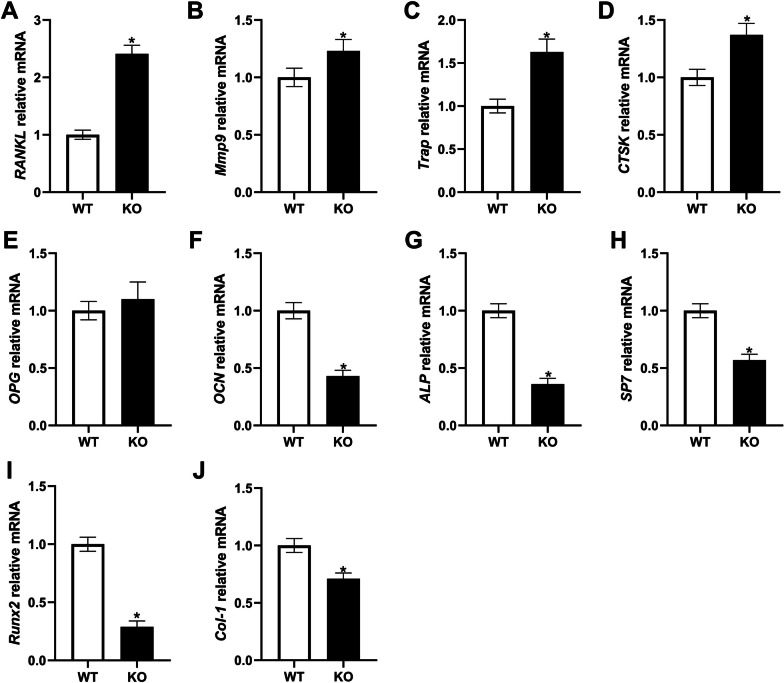
Results: In hepcidin1-knockout mice, the ferritin and iron contents in liver and tibia were significantly increased. Iron accumulation induced by hepcidin1 knockout caused a phenotype of low bone mass and deteriorated bone microarchitecture. Osteogenic marker was decreased and osteoclast marker was increased in mice, accompanied by increased oxidative stress level. The mRNA expression levels of osteoclast differentiation markers (RANKL, Mmp9, OPG, Trap, and CTSK) were up-regulated, while bone formation markers (OCN, ALP, Runx2, SP7, and Col-1) were down-regulated in model group, compared to wild type mice. In vitro, hepcidin1 knockdown inhibited proliferation and osteogenic differentiation, while promoted apoptosis, with increased levels of iron and ferritin.

Conclusion: Iron accumulation induced by hepcidin1 deficiency aggravates the progression of aging osteoporosis via inhibiting osteogenesis and promoting osteoclast genesis.

Keywords: Aged mice; Hepcidin; Iron; Osteoporosis.


Conflict of interest statement

The authors declare that they have no competing interest.

Figures

Fig. 1
Fig. 1
General and iron parameters in hepcidin1 knockout (KO) KO and wild type (WT) male mice at the age of 18 months. A body weight. B serum hepcidin level. C serum ferritin level. D liver iron concentration. E liver iron concentration. The bar graph shows the means ± standard deviation (SD). *P < 0.05 vs. WT group
Fig. 2
Fig. 2
KO mice exhibit iron overload. Representative liver sections stained with Perls Prussian blue for iron shows predominantly periportal accumulation of iron of KO mice. Original magnification ×400 (black arrow is portal vein, red arrow is iron deposition)
Fig. 3
Fig. 3
Mouse femoral metaphyseal trabecular and cortical bone architecture examined by Micro-CT
Fig. 4
Fig. 4
Evaluation of bone resorption, bone formation markers and oxidative stress. A Serum levels of the bone resorption markers C-terminal telopeptide of type 1 collagen. B markers of bone formation osteocalcin. C, D Levels of the oxidative stress markers superoxide dismutase and malondialdehyde. The bar graph shows the means ± SD. *P < 0.05 vs. WT group
Fig. 5
Fig. 5
Expression of genes related to bone formation and resorption. The mRNA expression levels of A RANKL. B Mmp9. C Trap. D CTSK. E OPG. F OCN. G ALP. H SP7. I Runx2. J Col-1. The bar graph shows the means ± SD. *P < 0.05 vs. WT group
Fig. 6
Fig. 6
A The cell viability of mBMSCs treated with different concentrations of D-galactose (0–100 g/L) for 24 h was measured by CCK-8. B The transfection efficiency of hepcidin1 in mBMSCs was detected by real-time quantitative PCR. C The effect of hepcidin1 on the viability of mBMSCs determined by CCK-8. D Apoptosis of mBMSCs was detected by flow cytometry. E The changes in calcium nodules after transfection of mBMSCs were evaluated by Alizarin red staining (Amplification: 200× , Scale: 100 μm). F The proliferation of mBMSCs was detected by EdU assay. G The concentration of iron ions in mBMSCs was detected by Iron Colorimetric Assay Kit. H The expression level of ferritin protein was detected by western blot. *P < 0.05, **P < 0.01, ***P < 0.001 vs. Control group; #P < 0.05, ##P < 0.01, ###P < 0.001 vs. D-gal + siNC group

References

    1. Conference CD. Diagnosis, prophylaxis and treatment of osteoporosis. Am J Med. 1993;94(6):646–650. doi: 10.1016/0002-9343(93)90218-E. - DOI - PubMed
    1. Blau DM, Weinberg BA. Why the US science and engineering workforce is aging rapidly. Proc Natl Acad Sci U S A. 2017;114(15):3879–3884. doi: 10.1073/pnas.1611748114. - DOI - PMC - PubMed
    1. Cheung WH, et al. Fracture healing in osteoporotic bone. Injury. 2016;47(Suppl 2):S21–S26. doi: 10.1016/S0020-1383(16)47004-X. - DOI - PubMed
    1. Johnell O, Kanis JA. An estimate of the worldwide prevalence and disability associated with osteoporotic fractures. Osteoporos Int. 2006;17(12):1726–1733. doi: 10.1007/s00198-006-0172-4. - DOI - PubMed
    1. Glaser DL, Kaplan FS. Osteoporosis. Definition and clinical presentation. Spine (Phila Pa 1976) 1997;22(24 Suppl):12s–16s. doi: 10.1097/00007632-199712151-00003. - DOI - PubMed